 Movie
Movie Controller


+ Open data

- Basic information
| Entry | Database: PDB / ID: 7mjd | |||||||||
|---|---|---|---|---|---|---|---|---|---|---|
| Title | Crystal Structure Analysis of ALDH1B1 | |||||||||
 Components Components | Aldehyde dehydrogenase X, mitochondrial | |||||||||
 Keywords Keywords | OXIDOREDUCTASE / mitochondrial metabolism / pancreas cancer / colorectal cancer | |||||||||
| Function / homology |  Function and homology information Function and homology informationethanol catabolic process / Ethanol oxidation / aldehyde dehydrogenase (NAD+) / aldehyde dehydrogenase (NAD+) activity / Mitochondrial protein degradation / NAD binding / carbohydrate metabolic process / mitochondrial matrix / mitochondrion / nucleoplasm / cytosol Similarity search - Function | |||||||||
| Biological species |  Homo sapiens (human) Homo sapiens (human) | |||||||||
| Method |  X-RAY DIFFRACTION / SYNCHROTRON / MOLECULAR REPLACEMENT / molecular replacement / Resolution: 2.12 Å X-RAY DIFFRACTION / SYNCHROTRON / MOLECULAR REPLACEMENT / molecular replacement / Resolution: 2.12 Å | |||||||||
 Authors Authors | Fernandez, D. / Chen, J.K. | |||||||||
| Funding support |  United States, 2items United States, 2items
| |||||||||
 Citation Citation | Journal: Nat.Chem.Biol. / Year: 2022 Title: Inhibitors targeted to aldehyde dehydrogenase Authors: Fernandez, D. | |||||||||
| History |
|
- Structure visualization
| Structure viewer | Molecule: MolmilJmol/JSmol |
|---|
- Downloads & links
-Download
| PDBx/mmCIF format | 7mjd.cif.gz | 214.7 KB | Display | PDBx/mmCIF format |
|---|---|---|---|---|
| PDB format | pdb7mjd.ent.gz | 167 KB | Display | PDB format |
| PDBx/mmJSON format | 7mjd.json.gz | Tree view | PDBx/mmJSON format | |
| Others |  Other downloads Other downloads |
-Validation report
| Summary document | 7mjd_validation.pdf.gz | 1.6 MB | Display | wwPDB validaton report |
|---|---|---|---|---|
| Full document | 7mjd_full_validation.pdf.gz | 1.6 MB | Display | |
| Data in XML | 7mjd_validation.xml.gz | 38.6 KB | Display | |
| Data in CIF | 7mjd_validation.cif.gz | 53.1 KB | Display | |
| Arichive directory | https://data.pdbj.org/pub/pdb/validation_reports/mj/7mjdftp://data.pdbj.org/pub/pdb/validation_reports/mj/7mjd | HTTPS FTP |
-Related structure data
| Related structure data | 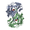 7mjcC 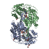 7radC 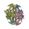 3n80S S: Starting model for refinement C: citing same article ( |
|---|---|
| Similar structure data |
-Links
 PDBj
PDBj


- Assembly
| Deposited unit | 
| |||||||||
|---|---|---|---|---|---|---|---|---|---|---|
| 1 |
| |||||||||
| Unit cell |
| |||||||||
| Components on special symmetry positions |
|
-Components
-Protein , 1 types, 2 molecules AB
| #1: Protein | Mass: 55101.434 Da / Num. of mol.: 2 Source method: isolated from a genetically manipulated source Source: (gene. exp.) Homo sapiens (human) / Gene: ALDH1B1, ALDH5, ALDHX / Production host:  |
|---|
-Non-polymers , 6 types, 212 molecules 


| #2: Chemical |  Mass: 22.990 Da / Num. of mol.: 2 / Source method: obtained synthetically / Formula: Na Mass: 22.990 Da / Num. of mol.: 2 / Source method: obtained synthetically / Formula: Na#3: Chemical |  Mass: 663.425 Da / Num. of mol.: 2 / Source method: obtained synthetically / Formula: C21H27N7O14P2 / Comment: NAD*YM Mass: 663.425 Da / Num. of mol.: 2 / Source method: obtained synthetically / Formula: C21H27N7O14P2 / Comment: NAD*YM#4: Chemical |  Mass: 395.516 Da / Num. of mol.: 2 / Source method: obtained synthetically / Formula: C27H27N2O / Feature type: SUBJECT OF INVESTIGATION Mass: 395.516 Da / Num. of mol.: 2 / Source method: obtained synthetically / Formula: C27H27N2O / Feature type: SUBJECT OF INVESTIGATION#5: Chemical | ChemComp-1PE /  Mass: 238.278 Da / Num. of mol.: 18 / Source method: obtained synthetically / Formula: C10H22O6 / Comment: precipitant*YM Mass: 238.278 Da / Num. of mol.: 18 / Source method: obtained synthetically / Formula: C10H22O6 / Comment: precipitant*YM#6: Chemical |  Mass: 92.094 Da / Num. of mol.: 2 / Source method: obtained synthetically / Formula: C3H8O3 Mass: 92.094 Da / Num. of mol.: 2 / Source method: obtained synthetically / Formula: C3H8O3#7: Water | ChemComp-HOH / | Mass: 18.015 Da / Num. of mol.: 186 / Source method: isolated from a natural source / Formula: H2O |
|---|
-Details
| Has ligand of interest | Y |
|---|
-Experimental details
-Experiment
| Experiment | Method: X-RAY DIFFRACTION / Number of used crystals: 1 |
|---|
- Sample preparation
| Crystal | Density Matthews: 2.49 Å3/Da / Density % sol: 51 % |
|---|---|
| Crystal grow | Temperature: 289 K / Method: vapor diffusion, sitting drop / pH: 8.5 / Details: PEG 4000, glycerol, tris-bicine, ethyleneglycols |
-Data collection
| Diffraction | Mean temperature: 100 K / Serial crystal experiment: N | |||||||||||||||||||||||||||||||||||||||||||||||||||||||||||||||||||||||||||||||||||||||||||||||||||||||||||||||||||||||||
|---|---|---|---|---|---|---|---|---|---|---|---|---|---|---|---|---|---|---|---|---|---|---|---|---|---|---|---|---|---|---|---|---|---|---|---|---|---|---|---|---|---|---|---|---|---|---|---|---|---|---|---|---|---|---|---|---|---|---|---|---|---|---|---|---|---|---|---|---|---|---|---|---|---|---|---|---|---|---|---|---|---|---|---|---|---|---|---|---|---|---|---|---|---|---|---|---|---|---|---|---|---|---|---|---|---|---|---|---|---|---|---|---|---|---|---|---|---|---|---|---|---|---|
| Diffraction source | Source: SYNCHROTRON / Site: SSRL / Beamline: BL12-2 / Wavelength: 0.97946 Å | |||||||||||||||||||||||||||||||||||||||||||||||||||||||||||||||||||||||||||||||||||||||||||||||||||||||||||||||||||||||||
| Detector | Type: DECTRIS PILATUS 6M / Detector: PIXEL / Date: Dec 16, 2018 Details: Rh coated collimating mirrors, K-B focusing mirrors | |||||||||||||||||||||||||||||||||||||||||||||||||||||||||||||||||||||||||||||||||||||||||||||||||||||||||||||||||||||||||
| Radiation | Monochromator: Liquid nitrogen-cooled double crystal, non fixed exit slit Protocol: SINGLE WAVELENGTH / Monochromatic (M) / Laue (L): M / Scattering type: x-ray | |||||||||||||||||||||||||||||||||||||||||||||||||||||||||||||||||||||||||||||||||||||||||||||||||||||||||||||||||||||||||
| Radiation wavelength | Wavelength: 0.97946 Å / Relative weight: 1 | |||||||||||||||||||||||||||||||||||||||||||||||||||||||||||||||||||||||||||||||||||||||||||||||||||||||||||||||||||||||||
| Reflection | Resolution: 2.12→88.369 Å / Num. all: 64238 / Num. obs: 64238 / % possible obs: 99.9 % / Redundancy: 6.7 % / Rpim(I) all: 0.034 / Rrim(I) all: 0.088 / Rsym value: 0.081 / Net I/av σ(I): 3.8 / Net I/σ(I): 11.4 / Num. measured all: 431121 | |||||||||||||||||||||||||||||||||||||||||||||||||||||||||||||||||||||||||||||||||||||||||||||||||||||||||||||||||||||||||
| Reflection shell | Diffraction-ID: 1
|
-Phasing
| Phasing | Method: molecular replacement | |||||||||
|---|---|---|---|---|---|---|---|---|---|---|
| Phasing MR | Model details: Phaser MODE: MR_AUTO
|
- Processing
| Software |
| ||||||||||||||||||||||||||||||||||||||||||||||||||||||||||||
|---|---|---|---|---|---|---|---|---|---|---|---|---|---|---|---|---|---|---|---|---|---|---|---|---|---|---|---|---|---|---|---|---|---|---|---|---|---|---|---|---|---|---|---|---|---|---|---|---|---|---|---|---|---|---|---|---|---|---|---|---|---|
| Refinement | Method to determine structure: MOLECULAR REPLACEMENT Starting model: 3N80 Resolution: 2.12→30.37 Å / Cor.coef. Fo:Fc: 0.963 / Cor.coef. Fo:Fc free: 0.949 / SU B: 5.3 / SU ML: 0.137 / SU R Cruickshank DPI: 0.2089 / Cross valid method: THROUGHOUT / σ(F): 0 / ESU R: 0.209 / ESU R Free: 0.173 / Stereochemistry target values: MAXIMUM LIKELIHOOD Details: HYDROGENS HAVE BEEN ADDED IN THE RIDING POSITIONS U VALUES : REFINED INDIVIDUALLY
| ||||||||||||||||||||||||||||||||||||||||||||||||||||||||||||
| Solvent computation | Ion probe radii: 0.8 Å / Shrinkage radii: 0.8 Å / VDW probe radii: 1.2 Å / Solvent model: MASK | ||||||||||||||||||||||||||||||||||||||||||||||||||||||||||||
| Displacement parameters | Biso max: 106.71 Å2 / Biso mean: 48.122 Å2 / Biso min: 23.77 Å2
| ||||||||||||||||||||||||||||||||||||||||||||||||||||||||||||
| Refinement step | Cycle: final / Resolution: 2.12→30.37 Å
| ||||||||||||||||||||||||||||||||||||||||||||||||||||||||||||
| Refine LS restraints |
| ||||||||||||||||||||||||||||||||||||||||||||||||||||||||||||
| LS refinement shell | Resolution: 2.12→2.175 Å / Rfactor Rfree error: 0 / Total num. of bins used: 20
|
