[English] 日本語
 Yorodumi
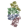
- PDB-7miy: Human N-type voltage-gated calcium channel Cav2.2 at 3.1 Angstrom... -
+ Open data
- Basic information
| Entry | Database: PDB / ID: 7miy | |||||||||||||||||||||||||||||||||||||||
|---|---|---|---|---|---|---|---|---|---|---|---|---|---|---|---|---|---|---|---|---|---|---|---|---|---|---|---|---|---|---|---|---|---|---|---|---|---|---|---|---|
| Title | Human N-type voltage-gated calcium channel Cav2.2 at 3.1 Angstrom resolution | |||||||||||||||||||||||||||||||||||||||
 Components Components | (Voltage-dependent ...) x 3 | |||||||||||||||||||||||||||||||||||||||
 Keywords Keywords | TRANSPORT PROTEIN / Cav2.2 / Channels / Calcium Ion-Selective | |||||||||||||||||||||||||||||||||||||||
| Function / homology |  Function and homology information Function and homology informationpositive regulation of high voltage-gated calcium channel activity / regulation of membrane repolarization during action potential / Presynaptic depolarization and calcium channel opening / positive regulation of calcium ion-dependent exocytosis of neurotransmitter / calcium ion transmembrane transport via high voltage-gated calcium channel / high voltage-gated calcium channel activity / membrane depolarization during bundle of His cell action potential / L-type voltage-gated calcium channel complex / regulation of ventricular cardiac muscle cell membrane repolarization / NCAM1 interactions ...positive regulation of high voltage-gated calcium channel activity / regulation of membrane repolarization during action potential / Presynaptic depolarization and calcium channel opening / positive regulation of calcium ion-dependent exocytosis of neurotransmitter / calcium ion transmembrane transport via high voltage-gated calcium channel / high voltage-gated calcium channel activity / membrane depolarization during bundle of His cell action potential / L-type voltage-gated calcium channel complex / regulation of ventricular cardiac muscle cell membrane repolarization / NCAM1 interactions / cardiac muscle cell action potential involved in contraction / calcium ion transport into cytosol / regulation of calcium ion transmembrane transport via high voltage-gated calcium channel / voltage-gated calcium channel complex / Mechanical load activates signaling by PIEZO1 and integrins in osteocytes / response to amyloid-beta / regulation of heart rate by cardiac conduction / regulation of calcium ion transport / calcium ion import across plasma membrane / neuronal dense core vesicle / voltage-gated calcium channel activity / presynaptic active zone membrane / sarcoplasmic reticulum / protein localization to plasma membrane / calcium channel regulator activity / Regulation of insulin secretion / modulation of chemical synaptic transmission / GABA-ergic synapse / cellular response to amyloid-beta / Adrenaline,noradrenaline inhibits insulin secretion / calcium ion transport / T cell receptor signaling pathway / amyloid-beta binding / chemical synaptic transmission / neuronal cell body / synapse / calcium ion binding / extracellular exosome / ATP binding / metal ion binding / membrane / plasma membrane / cytosol Similarity search - Function | |||||||||||||||||||||||||||||||||||||||
| Biological species |  Homo sapiens (human) Homo sapiens (human) | |||||||||||||||||||||||||||||||||||||||
| Method | ELECTRON MICROSCOPY / single particle reconstruction / cryo EM / Resolution: 3.1 Å | |||||||||||||||||||||||||||||||||||||||
 Authors Authors | Yan, N. / Gao, S. / Yao, X. | |||||||||||||||||||||||||||||||||||||||
| Funding support |  United States, 1items United States, 1items
| |||||||||||||||||||||||||||||||||||||||
 Citation Citation |  Journal: Nature / Year: 2021 Journal: Nature / Year: 2021Title: Structure of human Ca2.2 channel blocked by the painkiller ziconotide. Authors: Shuai Gao / Xia Yao / Nieng Yan /  Abstract: The neuronal-type (N-type) voltage-gated calcium (Ca) channels, which are designated Ca2.2, have an important role in the release of neurotransmitters. Ziconotide is a Ca2.2-specific peptide pore ...The neuronal-type (N-type) voltage-gated calcium (Ca) channels, which are designated Ca2.2, have an important role in the release of neurotransmitters. Ziconotide is a Ca2.2-specific peptide pore blocker that has been clinically used for treating intractable pain. Here we present cryo-electron microscopy structures of human Ca2.2 (comprising the core α1 and the ancillary α2δ-1 and β3 subunits) in the presence or absence of ziconotide. Ziconotide is thoroughly coordinated by helices P1 and P2, which support the selectivity filter, and the extracellular loops (ECLs) in repeats II, III and IV of α1. To accommodate ziconotide, the ECL of repeat III and α2δ-1 have to tilt upward concertedly. Three of the voltage-sensing domains (VSDs) are in a depolarized state, whereas the VSD of repeat II exhibits a down conformation that is stabilized by Ca2-unique intracellular segments and a phosphatidylinositol 4,5-bisphosphate molecule. Our studies reveal the molecular basis for Ca2.2-specific pore blocking by ziconotide and establish the framework for investigating electromechanical coupling in Ca channels. | |||||||||||||||||||||||||||||||||||||||
| History |
|
- Structure visualization
| Movie |
 Movie viewer Movie viewer |
|---|---|
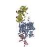
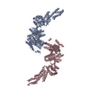
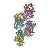
| Structure viewer | Molecule:  Molmil Molmil Jmol/JSmol Jmol/JSmol |
- Downloads & links
- Download
| PDBx/mmCIF format |  7miy.cif.gz 7miy.cif.gz | 515 KB | Display |  PDBx/mmCIF format PDBx/mmCIF format |
|---|---|---|---|---|
| PDB format |  pdb7miy.ent.gz pdb7miy.ent.gz | 398.9 KB | Display |  PDB format PDB format |
| PDBx/mmJSON format |  7miy.json.gz 7miy.json.gz | Tree view |  PDBx/mmJSON format PDBx/mmJSON format | |
| Others |  Other downloads Other downloads |
-Validation report
| Summary document |  7miy_validation.pdf.gz 7miy_validation.pdf.gz | 1.8 MB | Display |  wwPDB validaton report wwPDB validaton report |
|---|---|---|---|---|
| Full document |  7miy_full_validation.pdf.gz 7miy_full_validation.pdf.gz | 1.9 MB | Display | |
| Data in XML |  7miy_validation.xml.gz 7miy_validation.xml.gz | 77.9 KB | Display | |
| Data in CIF |  7miy_validation.cif.gz 7miy_validation.cif.gz | 115.5 KB | Display | |
| Arichive directory |  https://data.pdbj.org/pub/pdb/validation_reports/mi/7miy https://data.pdbj.org/pub/pdb/validation_reports/mi/7miy ftp://data.pdbj.org/pub/pdb/validation_reports/mi/7miy ftp://data.pdbj.org/pub/pdb/validation_reports/mi/7miy | HTTPS FTP |
-Related structure data
| Related structure data |  23868MC  7mixC M: map data used to model this data C: citing same article ( |
|---|---|
| Similar structure data |
- Links
Links
- Assembly
| Deposited unit | 
|
|---|---|
| 1 |
|
- Components
-Voltage-dependent ... , 3 types, 3 molecules ACD
| #1: Protein | Mass: 262831.781 Da / Num. of mol.: 1 Source method: isolated from a genetically manipulated source Source: (gene. exp.)  Homo sapiens (human) / Gene: CACNA1B, CACH5, CACNL1A5 / Production host: Homo sapiens (human) / Gene: CACNA1B, CACH5, CACNL1A5 / Production host:  Homo sapiens (human) / References: UniProt: Q00975 Homo sapiens (human) / References: UniProt: Q00975 |
|---|---|
| #2: Protein | Mass: 54607.852 Da / Num. of mol.: 1 Source method: isolated from a genetically manipulated source Source: (gene. exp.)  Homo sapiens (human) / Gene: CACNB3, CACNLB3 / Production host: Homo sapiens (human) / Gene: CACNB3, CACNLB3 / Production host:  Homo sapiens (human) / References: UniProt: P54284 Homo sapiens (human) / References: UniProt: P54284 |
| #3: Protein | Mass: 124692.469 Da / Num. of mol.: 1 Source method: isolated from a genetically manipulated source Source: (gene. exp.)  Homo sapiens (human) / Gene: CACNA2D1, CACNL2A, CCHL2A, MHS3 / Production host: Homo sapiens (human) / Gene: CACNA2D1, CACNL2A, CCHL2A, MHS3 / Production host:  Homo sapiens (human) / References: UniProt: P54289 Homo sapiens (human) / References: UniProt: P54289 |
-Sugars , 4 types, 8 molecules 
| #4: Polysaccharide | 2-acetamido-2-deoxy-beta-D-glucopyranose-(1-4)-2-acetamido-2-deoxy-beta-D-glucopyranose-(1-4)-2- ...2-acetamido-2-deoxy-beta-D-glucopyranose-(1-4)-2-acetamido-2-deoxy-beta-D-glucopyranose-(1-4)-2-acetamido-2-deoxy-beta-D-glucopyranose Source method: isolated from a genetically manipulated source | ||||
|---|---|---|---|---|---|
| #5: Polysaccharide | Source method: isolated from a genetically manipulated source #6: Polysaccharide | 2-acetamido-2-deoxy-beta-D-glucopyranose-(1-4)-2-acetamido-2-deoxy-beta-D-glucopyranose-(1-4)-2- ...2-acetamido-2-deoxy-beta-D-glucopyranose-(1-4)-2-acetamido-2-deoxy-beta-D-glucopyranose-(1-4)-2-acetamido-2-deoxy-beta-D-glucopyranose-(1-4)-2-acetamido-2-deoxy-beta-D-glucopyranose | Source method: isolated from a genetically manipulated source #7: Sugar | |
-Non-polymers , 5 types, 12 molecules 


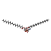


| #8: Chemical | | #9: Chemical | #10: Chemical | ChemComp-CLR / #11: Chemical | #12: Chemical | ChemComp-PT5 / [( | |
|---|
-Details
| Has ligand of interest | Y |
|---|---|
| Has protein modification | Y |
-Experimental details
-Experiment
| Experiment | Method: ELECTRON MICROSCOPY |
|---|---|
| EM experiment | Aggregation state: PARTICLE / 3D reconstruction method: single particle reconstruction |
- Sample preparation
| Component | Name: Cav2.2 / Type: COMPLEX / Entity ID: #1-#3 / Source: RECOMBINANT |
|---|---|
| Molecular weight | Experimental value: NO |
| Source (natural) | Organism:  Homo sapiens (human) Homo sapiens (human) |
| Source (recombinant) | Organism:  Homo sapiens (human) Homo sapiens (human) |
| Buffer solution | pH: 7.4 |
| Specimen | Conc.: 20 mg/ml / Embedding applied: NO / Shadowing applied: NO / Staining applied: NO / Vitrification applied: YES / Details: Cav2.2 |
| Vitrification | Instrument: FEI VITROBOT MARK IV / Cryogen name: ETHANE / Humidity: 100 % / Chamber temperature: 281 K / Details: blot for 6 seconds before plunging |
- Electron microscopy imaging
| Experimental equipment |  Model: Titan Krios / Image courtesy: FEI Company |
|---|---|
| Microscopy | Model: FEI TITAN KRIOS |
| Electron gun | Electron source:  FIELD EMISSION GUN / Accelerating voltage: 300 kV / Illumination mode: FLOOD BEAM FIELD EMISSION GUN / Accelerating voltage: 300 kV / Illumination mode: FLOOD BEAM |
| Electron lens | Mode: BRIGHT FIELD |
| Image recording | Electron dose: 50 e/Å2 / Film or detector model: GATAN K2 SUMMIT (4k x 4k) |
- Processing
| Software | Name: PHENIX / Version: 1.19.2_4158: / Classification: refinement | ||||||||||||||||||||||||
|---|---|---|---|---|---|---|---|---|---|---|---|---|---|---|---|---|---|---|---|---|---|---|---|---|---|
| EM software | Name: PHENIX / Category: model refinement | ||||||||||||||||||||||||
| CTF correction | Type: PHASE FLIPPING AND AMPLITUDE CORRECTION | ||||||||||||||||||||||||
| 3D reconstruction | Resolution: 3.1 Å / Resolution method: FSC 0.143 CUT-OFF / Num. of particles: 56615 / Symmetry type: POINT | ||||||||||||||||||||||||
| Refine LS restraints |
|
 Movie
Movie Controller





 PDBj
PDBj


