 Movie
Movie Controller

[English] 日本語
 Yorodumi
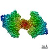
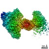
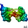
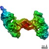
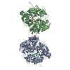
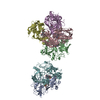
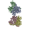
- PDB-7mi9: Full integration complex of Cas1/Cas2 from Cas4-containing system -
+ Open data

- Basic information
| Entry | Database: PDB / ID: 7mi9 | ||||||
|---|---|---|---|---|---|---|---|
| Title | Full integration complex of Cas1/Cas2 from Cas4-containing system | ||||||
 Components Components |
| ||||||
 Keywords Keywords | HYDROLASE/DNA /  CRISPR/Cas / Cas4 / PAM recognition / full integration / HYDROLASE-DNA complex CRISPR/Cas / Cas4 / PAM recognition / full integration / HYDROLASE-DNA complex | ||||||
| Function / homology |  Function and homology information Function and homology information5' to 3' exodeoxyribonuclease (nucleoside 3'-phosphate-forming) / exonuclease activity / maintenance of CRISPR repeat elements / RNA endonuclease activity / 4 iron, 4 sulfur cluster binding / endonuclease activity / defense response to virus / Hydrolases; Acting on ester bonds / DNA binding / metal ion bindingSimilarity search - Function | ||||||
| Biological species |  Geobacter sulfurreducens (bacteria) Geobacter sulfurreducens (bacteria) | ||||||
| Method | ELECTRON MICROSCOPY / single particle reconstruction / cryo EM / Resolution: 3.89 Å | ||||||
 Authors Authors | Hu, C.Y. / Ke, A.K. | ||||||
 Citation Citation | Journal: Nature / Year: 2021 Title: Mechanism for Cas4-assisted directional spacer acquisition in CRISPR-Cas. Authors: Chunyi Hu / Cristóbal Almendros / Ki Hyun Nam / Ana Rita Costa / Jochem N A Vink / Anna C Haagsma / Saket R Bagde / Stan J J Brouns / Ailong Ke /    Abstract: Prokaryotes adapt to challenges from mobile genetic elements by integrating spacers derived from foreign DNA in the CRISPR array. Spacer insertion is carried out by the Cas1-Cas2 integrase complex. A ...Prokaryotes adapt to challenges from mobile genetic elements by integrating spacers derived from foreign DNA in the CRISPR array. Spacer insertion is carried out by the Cas1-Cas2 integrase complex. A substantial fraction of CRISPR-Cas systems use a Fe-S cluster containing Cas4 nuclease to ensure that spacers are acquired from DNA flanked by a protospacer adjacent motif (PAM) and inserted into the CRISPR array unidirectionally, so that the transcribed CRISPR RNA can guide target searching in a PAM-dependent manner. Here we provide a high-resolution mechanistic explanation for the Cas4-assisted PAM selection, spacer biogenesis and directional integration by type I-G CRISPR in Geobacter sulfurreducens, in which Cas4 is naturally fused with Cas1, forming Cas4/Cas1. During biogenesis, only DNA duplexes possessing a PAM-embedded 3'-overhang trigger Cas4/Cas1-Cas2 assembly. During this process, the PAM overhang is specifically recognized and sequestered, but is not cleaved by Cas4. This 'molecular constipation' prevents the PAM-side prespacer from participating in integration. Lacking such sequestration, the non-PAM overhang is trimmed by host nucleases and integrated to the leader-side CRISPR repeat. Half-integration subsequently triggers PAM cleavage and Cas4 dissociation, allowing spacer-side integration. Overall, the intricate molecular interaction between Cas4 and Cas1-Cas2 selects PAM-containing prespacers for integration and couples the timing of PAM processing with the stepwise integration to establish directionality. | ||||||
| History |
|
- Structure visualization
| Movie |
Movie viewer |
|---|---|
| Structure viewer | Molecule: MolmilJmol/JSmol |
- Downloads & links
-Download
| PDBx/mmCIF format | 7mi9.cif.gz | 365.9 KB | Display | PDBx/mmCIF format |
|---|---|---|---|---|
| PDB format | pdb7mi9.ent.gz | 288.7 KB | Display | PDB format |
| PDBx/mmJSON format | 7mi9.json.gz | Tree view | PDBx/mmJSON format | |
| Others |  Other downloads Other downloads |
-Validation report
| Arichive directory | https://data.pdbj.org/pub/pdb/validation_reports/mi/7mi9ftp://data.pdbj.org/pub/pdb/validation_reports/mi/7mi9 | HTTPS FTP |
|---|
-Related structure data
| Related structure data |  23843MC 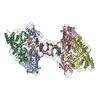 7mi4C 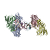 7mi5C 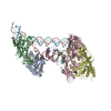 7mibC 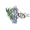 7midC C: citing same article ( M: map data used to model this data |
|---|---|
| Similar structure data |


-Links
 PDBj
PDBj


- Assembly
| Deposited unit | 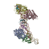
|
|---|---|
| 1 |
|
-Components
-CRISPR-associated ... , 2 types, 6 molecules ABCDEF
| #1: Protein | Mass: 62598.496 Da / Num. of mol.: 4 Source method: isolated from a genetically manipulated source Source: (gene. exp.) Geobacter sulfurreducens (bacteria) / Strain: ATCC 51573 / DSM 12127 / PCA / Gene: cas4-cas1, GSU0057 / Production host: Escherichia coli (E. coli)References: UniProt: Q74H36, Hydrolases; Acting on ester bonds, 5' to 3' exodeoxyribonuclease (nucleoside 3'-phosphate-forming)#2: Protein | Mass: 11190.176 Da / Num. of mol.: 2 Source method: isolated from a genetically manipulated source Source: (gene. exp.) Geobacter sulfurreducens (bacteria) / Strain: ATCC 51573 / DSM 12127 / PCA / Gene: cas2, GSU0058 / Production host: Escherichia coli (E. coli)References: UniProt: Q74H35, Hydrolases; Acting on ester bonds |
|---|
-DNA chain , 4 types, 4 molecules GHIJ
| #3: DNA chain | Mass: 24664.730 Da / Num. of mol.: 1 / Source method: obtained synthetically / Source: (synth.) Geobacter sulfurreducens (bacteria) |
|---|---|
| #4: DNA chain | Mass: 22123.074 Da / Num. of mol.: 1 / Source method: obtained synthetically / Source: (synth.) Geobacter sulfurreducens (bacteria) |
| #5: DNA chain | Mass: 3705.445 Da / Num. of mol.: 1 / Source method: obtained synthetically / Source: (synth.) Geobacter sulfurreducens (bacteria) |
| #6: DNA chain | Mass: 1818.231 Da / Num. of mol.: 1 / Source method: obtained synthetically / Source: (synth.) Geobacter sulfurreducens (bacteria) |
-Experimental details
-Experiment
| Experiment | Method: ELECTRON MICROSCOPY |
|---|---|
| EM experiment | Aggregation state: PARTICLE / 3D reconstruction method: single particle reconstruction |
- Sample preparation
| Component |
| |||||||||||||||||||||||||||||||||||
|---|---|---|---|---|---|---|---|---|---|---|---|---|---|---|---|---|---|---|---|---|---|---|---|---|---|---|---|---|---|---|---|---|---|---|---|---|
| Molecular weight |
| |||||||||||||||||||||||||||||||||||
| Source (natural) | Organism: Geobacter sulfurreducens (bacteria) | |||||||||||||||||||||||||||||||||||
| Source (recombinant) | Organism: Escherichia coli (E. coli) | |||||||||||||||||||||||||||||||||||
| Buffer solution | pH: 7.5 / Details: WITH 5 mM DTT | |||||||||||||||||||||||||||||||||||
| Buffer component | Conc.: 150 mM / Name: sodium chloride / Formula: NaClSodium chloride | |||||||||||||||||||||||||||||||||||
| Specimen | Conc.: 1 mg/ml / Embedding applied: NO / Shadowing applied: NO / Staining applied: NO / Vitrification applied: YES | |||||||||||||||||||||||||||||||||||
| Specimen support | Grid type: Quantifoil R1.2/1.3 | |||||||||||||||||||||||||||||||||||
| Vitrification | Instrument: FEI VITROBOT MARK IV / Cryogen name: ETHANE / Humidity: 100 % / Chamber temperature: 298 K / Details: 6 seconds |
- Electron microscopy imaging
| Microscopy | Model: TFS TALOS |
|---|---|
| Electron gun | Electron source: FIELD EMISSION GUN / Accelerating voltage: 200 kV / Illumination mode: FLOOD BEAM |
| Electron lens | Mode: DIFFRACTION / Nominal defocus min: 1500 nm / Cs: 2.7 mm / C2 aperture diameter: 100 µm |
| Image recording | Average exposure time: 0.35 sec. / Electron dose: 50 e/Å2 / Film or detector model: GATAN K3 BIOQUANTUM (6k x 4k) / Num. of real images: 1200 |
| EM imaging optics | Phase plate: VOLTA PHASE PLATE |
- Processing
| Software | Name: PHENIX / Version: 1.18.2_3874: / Classification: refinement | ||||||||||||||||||||||||
|---|---|---|---|---|---|---|---|---|---|---|---|---|---|---|---|---|---|---|---|---|---|---|---|---|---|
| EM software |
| ||||||||||||||||||||||||
| CTF correction | Type: PHASE FLIPPING AND AMPLITUDE CORRECTION | ||||||||||||||||||||||||
| 3D reconstruction | Resolution: 3.89 Å / Resolution method: FSC 0.143 CUT-OFF / Num. of particles: 80000 / Symmetry type: POINT | ||||||||||||||||||||||||
| Atomic model building | Protocol: OTHER | ||||||||||||||||||||||||
| Refine LS restraints |
|
