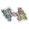
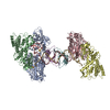
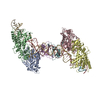
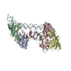
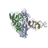
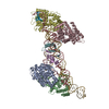
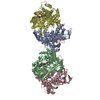
Journal: Nature / Year: 2021 Title: Mechanism for Cas4-assisted directional spacer acquisition in CRISPR-Cas. Authors: Chunyi Hu / Cristóbal Almendros / Ki Hyun Nam / Ana Rita Costa / Jochem N A Vink / Anna C Haagsma / Saket R Bagde / Stan J J Brouns / Ailong Ke / Abstract: Prokaryotes adapt to challenges from mobile genetic elements by integrating spacers derived from foreign DNA in the CRISPR array. Spacer insertion is carried out by the Cas1-Cas2 integrase complex. A ...Prokaryotes adapt to challenges from mobile genetic elements by integrating spacers derived from foreign DNA in the CRISPR array. Spacer insertion is carried out by the Cas1-Cas2 integrase complex. A substantial fraction of CRISPR-Cas systems use a Fe-S cluster containing Cas4 nuclease to ensure that spacers are acquired from DNA flanked by a protospacer adjacent motif (PAM) and inserted into the CRISPR array unidirectionally, so that the transcribed CRISPR RNA can guide target searching in a PAM-dependent manner. Here we provide a high-resolution mechanistic explanation for the Cas4-assisted PAM selection, spacer biogenesis and directional integration by type I-G CRISPR in Geobacter sulfurreducens, in which Cas4 is naturally fused with Cas1, forming Cas4/Cas1. During biogenesis, only DNA duplexes possessing a PAM-embedded 3'-overhang trigger Cas4/Cas1-Cas2 assembly. During this process, the PAM overhang is specifically recognized and sequestered, but is not cleaved by Cas4. This 'molecular constipation' prevents the PAM-side prespacer from participating in integration. Lacking such sequestration, the non-PAM overhang is trimmed by host nucleases and integrated to the leader-side CRISPR repeat. Half-integration subsequently triggers PAM cleavage and Cas4 dissociation, allowing spacer-side integration. Overall, the intricate molecular interaction between Cas4 and Cas1-Cas2 selects PAM-containing prespacers for integration and couples the timing of PAM processing with the stepwise integration to establish directionality.
History
Deposition
Apr 16, 2021
-
Header (metadata) release
Nov 17, 2021
-
Map release
Nov 17, 2021
-
Update
Nov 17, 2021
-
Current status
Nov 17, 2021
Processing site: RCSB / Status: Released
-
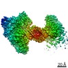
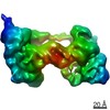
Structure visualization
Movie
Surface view with section colored by density value
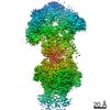
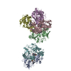
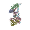
Entire : Half integration complex and Cas4 dissociated from PAM side
Entire
Name: Half integration complex and Cas4 dissociated from PAM side
Components
Complex: Half integration complex and Cas4 dissociated from PAM side
Protein or peptide: half integration complex of Cas1-Cas2
-
Supramolecule #1: Half integration complex and Cas4 dissociated from PAM side
Supramolecule
Name: Half integration complex and Cas4 dissociated from PAM side type: complex / ID: 1 / Parent: 0 / Macromolecule list: all / Details: half integration in cas4 containing system
In the structure databanks used in Yorodumi, some data are registered as the other names, "COVID-19 virus" and "2019-nCoV". Here are the details of the virus and the list of structure data.
Jan 31, 2019. EMDB accession codes are about to change! (news from PDBe EMDB page)
EMDB accession codes are about to change! (news from PDBe EMDB page)
The allocation of 4 digits for EMDB accession codes will soon come to an end. Whilst these codes will remain in use, new EMDB accession codes will include an additional digit and will expand incrementally as the available range of codes is exhausted. The current 4-digit format prefixed with “EMD-” (i.e. EMD-XXXX) will advance to a 5-digit format (i.e. EMD-XXXXX), and so on. It is currently estimated that the 4-digit codes will be depleted around Spring 2019, at which point the 5-digit format will come into force.
The EM Navigator/Yorodumi systems omit the EMD- prefix.
Related info.:Q: What is EMD? / ID/Accession-code notation in Yorodumi/EM Navigator
Yorodumi is a browser for structure data from EMDB, PDB, SASBDB, etc.
This page is also the successor to EM Navigator detail page, and also detail information page/front-end page for Omokage search.
The word "yorodu" (or yorozu) is an old Japanese word meaning "ten thousand". "mi" (miru) is to see.
Related info.:EMDB / PDB / SASBDB / Comparison of 3 databanks / Yorodumi Search / Aug 31, 2016. New EM Navigator & Yorodumi / Yorodumi Papers / Jmol/JSmol / Function and homology information / Changes in new EM Navigator and Yorodumi
 Movie
Movie Controller

 Yorodumi
 Open data
 Basic information
 Map data
 Sample
 Function and homology information
 Geobacter sulfurreducens (bacteria)
 Authors
 Citation


 Structure visualization
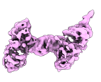
 Downloads & links
 emd_23849.png
 http://ftp.pdbj.org/pub/emdb/structures/EMD-23849
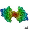


 Z (Sec.)
 Y (Row.)
 X (Col.)


 Sample components
 Processing
 Electron microscopy
 FIELD EMISSION GUN