 Movie
Movie Controller

[English] 日本語
 Yorodumi
- PDB-7mgv: Chryseobacterium gregarium RiPP-associated ATP-grasp ligase in co... -
+ Open data

- Basic information
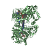
| Entry | Database: PDB / ID: 7mgv | ||||||
|---|---|---|---|---|---|---|---|
| Title | Chryseobacterium gregarium RiPP-associated ATP-grasp ligase in complex with ADP, and a leader and core peptide | ||||||
 Components Components |
| ||||||
 Keywords Keywords | LIGASE / Ribosomally synthesized / post-translationally modified peptide / Natural products ATP-grasp ligase / Precursor peptide / Graspetide omega-ester macrocycles macrolactone | ||||||
| Function / homology | ADENOSINE-5'-DIPHOSPHATE Function and homology information Function and homology information | ||||||
| Biological species |  Chryseobacterium gregarium DSM 19109 (bacteria) Chryseobacterium gregarium DSM 19109 (bacteria) | ||||||
| Method |  X-RAY DIFFRACTION / SYNCHROTRON / MOLECULAR REPLACEMENT / Resolution: 2.44 Å X-RAY DIFFRACTION / SYNCHROTRON / MOLECULAR REPLACEMENT / Resolution: 2.44 Å | ||||||
 Authors Authors | Bewley, C.A. / Zhao, G. / Kosek, D. / Dyda, F. | ||||||
 Citation Citation | Journal: J.Am.Chem.Soc. / Year: 2021 Title: Structural Basis for a Dual Function ATP Grasp Ligase That Installs Single and Bicyclic omega-Ester Macrocycles in a New Multicore RiPP Natural Product. Authors: Zhao, G. / Kosek, D. / Liu, H.B. / Ohlemacher, S.I. / Blackburne, B. / Nikolskaya, A. / Makarova, K.S. / Sun, J. / Barry Iii, C.E. / Koonin, E.V. / Dyda, F. / Bewley, C.A. | ||||||
| History |
|
- Structure visualization
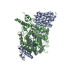
| Structure viewer | Molecule: MolmilJmol/JSmol |
|---|
- Downloads & links
-Download
| PDBx/mmCIF format | 7mgv.cif.gz | 195.5 KB | Display | PDBx/mmCIF format |
|---|---|---|---|---|
| PDB format | pdb7mgv.ent.gz | 124.5 KB | Display | PDB format |
| PDBx/mmJSON format | 7mgv.json.gz | Tree view | PDBx/mmJSON format | |
| Others |  Other downloads Other downloads |
-Validation report
| Arichive directory | https://data.pdbj.org/pub/pdb/validation_reports/mg/7mgvftp://data.pdbj.org/pub/pdb/validation_reports/mg/7mgv | HTTPS FTP |
|---|
-Related structure data
| Related structure data |  5ig9S S: Starting model for refinement |
|---|---|
| Similar structure data |


-Links
 PDBj
PDBj- Assembly
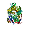
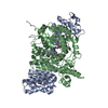
| Deposited unit | 
| ||||||||||||
|---|---|---|---|---|---|---|---|---|---|---|---|---|---|
| 1 |
| ||||||||||||
| Unit cell |
|
-Components
-Protein , 1 types, 2 molecules AB
| #1: Protein | Mass: 41624.074 Da / Num. of mol.: 2 Source method: isolated from a genetically manipulated source Source: (gene. exp.) Chryseobacterium gregarium DSM 19109 (bacteria)Production host: |
|---|
-Protein/peptide , 2 types, 3 molecules VUT
| #2: Protein/peptide | Mass: 1456.681 Da / Num. of mol.: 2 / Source method: obtained synthetically Source: (synth.) Chryseobacterium gregarium DSM 19109 (bacteria)#3: Protein/peptide | | Mass: 1212.242 Da / Num. of mol.: 1 / Source method: obtained synthetically Source: (synth.) Chryseobacterium gregarium DSM 19109 (bacteria) |
|---|
-Non-polymers , 3 types, 122 molecules 


| #4: Chemical |  Mass: 427.201 Da / Num. of mol.: 2 / Source method: obtained synthetically / Formula: C10H15N5O10P2 / Feature type: SUBJECT OF INVESTIGATION / Comment: ADP, energy-carrying molecule*YM Mass: 427.201 Da / Num. of mol.: 2 / Source method: obtained synthetically / Formula: C10H15N5O10P2 / Feature type: SUBJECT OF INVESTIGATION / Comment: ADP, energy-carrying molecule*YM#5: Chemical | ChemComp-GOL / |  Mass: 92.094 Da / Num. of mol.: 1 / Source method: obtained synthetically / Formula: C3H8O3 Mass: 92.094 Da / Num. of mol.: 1 / Source method: obtained synthetically / Formula: C3H8O3#6: Water | ChemComp-HOH / | Mass: 18.015 Da / Num. of mol.: 119 / Source method: isolated from a natural source / Formula: H2O |
|---|
-Details
| Has ligand of interest | Y |
|---|
-Experimental details
-Experiment
| Experiment | Method: X-RAY DIFFRACTION / Number of used crystals: 1 |
|---|
- Sample preparation
| Crystal | Density Matthews: 2.23 Å3/Da / Density % sol: 44.78 % |
|---|---|
| Crystal grow | Temperature: 293 K / Method: vapor diffusion, sitting drop / pH: 6.5 Details: 28% PEG 2000, 0.1 M bis-tris pH 6.5, 1 mM magnesium chloride |
-Data collection
| Diffraction | Mean temperature: 100 K / Serial crystal experiment: N |
|---|---|
| Diffraction source | Source: SYNCHROTRON / Site: APS  / Beamline: 22-ID / Wavelength: 0.96863 Å / Beamline: 22-ID / Wavelength: 0.96863 Å |
| Detector | Type: DECTRIS EIGER X 16M / Detector: PIXEL / Date: Nov 2, 2019 |
| Radiation | Protocol: SINGLE WAVELENGTH / Monochromatic (M) / Laue (L): M / Scattering type: x-ray |
| Radiation wavelength | Wavelength: 0.96863 Å / Relative weight: 1 |
| Reflection | Resolution: 2.44→30 Å / Num. obs: 29505 / % possible obs: 98.6 % / Redundancy: 6.2 % / Biso Wilson estimate: 47.39 Å2 / CC1/2: 0.993 / CC star: 0.998 / Rmerge(I) obs: 0.115 / Rpim(I) all: 0.044 / Rrim(I) all: 0.115 / Χ2: 0.894 / Net I/av σ(I): 16.8181 / Net I/σ(I): 18.5 |
| Reflection shell | Resolution: 2.44→2.48 Å / Redundancy: 3.4 % / Rmerge(I) obs: 0.554 / Mean I/σ(I) obs: 1.2 / Num. unique obs: 1438 / CC1/2: 0.687 / CC star: 0.903 / Rpim(I) all: 0.334 / Rrim(I) all: 0.652 / Χ2: 0.4 / % possible all: 97.4 |
- Processing
| Software |
| |||||||||||||||||||||||||||||||||||||||||||||||||||||||||||||||||||||||||||||||||||||||||||||||||||||||||
|---|---|---|---|---|---|---|---|---|---|---|---|---|---|---|---|---|---|---|---|---|---|---|---|---|---|---|---|---|---|---|---|---|---|---|---|---|---|---|---|---|---|---|---|---|---|---|---|---|---|---|---|---|---|---|---|---|---|---|---|---|---|---|---|---|---|---|---|---|---|---|---|---|---|---|---|---|---|---|---|---|---|---|---|---|---|---|---|---|---|---|---|---|---|---|---|---|---|---|---|---|---|---|---|---|---|---|
| Refinement | Method to determine structure: MOLECULAR REPLACEMENT Starting model: 5ig9 Resolution: 2.44→29.86 Å / SU ML: 0.3374 / Cross valid method: FREE R-VALUE / σ(F): 1.36 / Phase error: 26.0505 Stereochemistry target values: GeoStd + Monomer Library + CDL v1.2
| |||||||||||||||||||||||||||||||||||||||||||||||||||||||||||||||||||||||||||||||||||||||||||||||||||||||||
| Solvent computation | Shrinkage radii: 0.9 Å / VDW probe radii: 1.11 Å / Solvent model: FLAT BULK SOLVENT MODEL | |||||||||||||||||||||||||||||||||||||||||||||||||||||||||||||||||||||||||||||||||||||||||||||||||||||||||
| Displacement parameters | Biso mean: 52.86 Å2 | |||||||||||||||||||||||||||||||||||||||||||||||||||||||||||||||||||||||||||||||||||||||||||||||||||||||||
| Refinement step | Cycle: LAST / Resolution: 2.44→29.86 Å
| |||||||||||||||||||||||||||||||||||||||||||||||||||||||||||||||||||||||||||||||||||||||||||||||||||||||||
| Refine LS restraints |
| |||||||||||||||||||||||||||||||||||||||||||||||||||||||||||||||||||||||||||||||||||||||||||||||||||||||||
| LS refinement shell |
|
