 Movie
Movie Controller

[English] 日本語
 Yorodumi
- PDB-7md2: The F1 region of ammocidin-bound Saccharomyces cerevisiae ATP synthase -
+ Open data

- Basic information
| Entry | Database: PDB / ID: 7md2 | |||||||||||||||||||||||||||||||||||||||||||||||||||
|---|---|---|---|---|---|---|---|---|---|---|---|---|---|---|---|---|---|---|---|---|---|---|---|---|---|---|---|---|---|---|---|---|---|---|---|---|---|---|---|---|---|---|---|---|---|---|---|---|---|---|---|---|
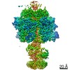
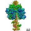
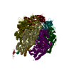
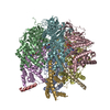
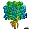
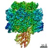
| Title | The F1 region of ammocidin-bound Saccharomyces cerevisiae ATP synthase | |||||||||||||||||||||||||||||||||||||||||||||||||||
 Components Components | (ATP synthase subunit ...) x 4 | |||||||||||||||||||||||||||||||||||||||||||||||||||
 Keywords Keywords | HYDROLASE/HYDROLASE INHIBITOR / F1-ATPase / inhibitor / anti-cancer / HYDROLASE / HYDROLASE-HYDROLASE INHIBITOR complex | |||||||||||||||||||||||||||||||||||||||||||||||||||
| Function / homology |  Function and homology information Function and homology information: / : / : / : / proton motive force-driven ATP synthesis / proton motive force-driven mitochondrial ATP synthesis / proton-transporting ATPase activity, rotational mechanism / H+-transporting two-sector ATPase / proton-transporting ATP synthase complex / proton-transporting ATP synthase activity, rotational mechanism ...: / : / : / : / proton motive force-driven ATP synthesis / proton motive force-driven mitochondrial ATP synthesis / proton-transporting ATPase activity, rotational mechanism / H+-transporting two-sector ATPase / proton-transporting ATP synthase complex / proton-transporting ATP synthase activity, rotational mechanism / ADP binding / mitochondrial inner membrane / ATP hydrolysis activity / mitochondrion / ATP binding Similarity search - Function | |||||||||||||||||||||||||||||||||||||||||||||||||||
| Biological species |  | |||||||||||||||||||||||||||||||||||||||||||||||||||
| Method | ELECTRON MICROSCOPY / single particle reconstruction / cryo EM / Resolution: 3.1 Å | |||||||||||||||||||||||||||||||||||||||||||||||||||
 Authors Authors | Guo, H. / Rubinstein, J.L. | |||||||||||||||||||||||||||||||||||||||||||||||||||
| Funding support |  Canada, Canada,  United States, 8items United States, 8items
| |||||||||||||||||||||||||||||||||||||||||||||||||||
 Citation Citation | Journal: Nat Chem Biol / Year: 2022 Title: Apoptolidin family glycomacrolides target leukemia through inhibition of ATP synthase. Authors: Benjamin J Reisman / Hui Guo / Haley E Ramsey / Madison T Wright / Bradley I Reinfeld / P Brent Ferrell / Gary A Sulikowski / W Kimryn Rathmell / Michael R Savona / Lars Plate / John L ...Authors: Benjamin J Reisman / Hui Guo / Haley E Ramsey / Madison T Wright / Bradley I Reinfeld / P Brent Ferrell / Gary A Sulikowski / W Kimryn Rathmell / Michael R Savona / Lars Plate / John L Rubinstein / Brian O Bachmann / Abstract: Cancer cells have long been recognized to exhibit unique bioenergetic requirements. The apoptolidin family of glycomacrolides are distinguished by their selective cytotoxicity towards oncogene- ...Cancer cells have long been recognized to exhibit unique bioenergetic requirements. The apoptolidin family of glycomacrolides are distinguished by their selective cytotoxicity towards oncogene-transformed cells, yet their molecular mechanism remains uncertain. We used photoaffinity analogs of the apoptolidins to identify the F subcomplex of mitochondrial ATP synthase as the target of apoptolidin A. Cryogenic electron microscopy (cryo-EM) of apoptolidin and ammocidin-ATP synthase complexes revealed a novel shared mode of inhibition that was confirmed by deep mutational scanning of the binding interface to reveal resistance mutations which were confirmed using CRISPR-Cas9. Ammocidin A was found to suppress leukemia progression in vivo at doses that were tolerated with minimal toxicity. The combination of cellular, structural, mutagenesis, and in vivo evidence defines the mechanism of action of apoptolidin family glycomacrolides and establishes a path to address oxidative phosphorylation-dependent cancers. | |||||||||||||||||||||||||||||||||||||||||||||||||||
| History |
|
- Structure visualization
| Movie |
Movie viewer |
|---|---|
| Structure viewer | Molecule: MolmilJmol/JSmol |
 UCSF Chimera
- Downloads & links
-Download
| PDBx/mmCIF format | 7md2.cif.gz | 1.1 MB | Display | PDBx/mmCIF format |
|---|---|---|---|---|
| PDB format | pdb7md2.ent.gz | Display | PDB format | |
| PDBx/mmJSON format | 7md2.json.gz | Tree view | PDBx/mmJSON format | |
| Others |  Other downloads Other downloads |
-Validation report
| Arichive directory | https://data.pdbj.org/pub/pdb/validation_reports/md/7md2ftp://data.pdbj.org/pub/pdb/validation_reports/md/7md2 | HTTPS FTP |
|---|
-Related structure data
| Related structure data | 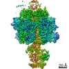 23763MC  7md3C M: map data used to model this data C: citing same article ( |
|---|---|
| Similar structure data |


-Links
 PDBj
PDBj


- Assembly
| Deposited unit | 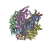
|
|---|---|
| 1 |
|
-Components
-ATP synthase subunit ... , 4 types, 8 molecules ABCDEFGH
| #1: Protein | Mass: 55007.402 Da / Num. of mol.: 3 / Source method: isolated from a natural source / Source: (natural) #2: Protein | Mass: 51181.082 Da / Num. of mol.: 3 / Source method: isolated from a natural source / Source: (natural) References: UniProt: A0A6A5PX46, H+-transporting two-sector ATPase #3: Protein | | Mass: 30657.160 Da / Num. of mol.: 1 / Source method: isolated from a natural source / Source: (natural) #4: Protein | | Mass: 6618.359 Da / Num. of mol.: 1 / Source method: isolated from a natural source / Source: (natural) |
|---|
-Non-polymers , 4 types, 8 molecules 


| #5: Chemical |  Mass: 507.181 Da / Num. of mol.: 3 / Source method: obtained synthetically / Formula: C10H16N5O13P3 / Comment: ATP, energy-carrying molecule*YM Mass: 507.181 Da / Num. of mol.: 3 / Source method: obtained synthetically / Formula: C10H16N5O13P3 / Comment: ATP, energy-carrying molecule*YM#6: Chemical |  Mass: 24.305 Da / Num. of mol.: 3 / Source method: obtained synthetically / Formula: Mg Mass: 24.305 Da / Num. of mol.: 3 / Source method: obtained synthetically / Formula: Mg#7: Chemical | ChemComp-PO4 / |  Mass: 94.971 Da / Num. of mol.: 1 / Source method: obtained synthetically / Formula: PO4 Mass: 94.971 Da / Num. of mol.: 1 / Source method: obtained synthetically / Formula: PO4#8: Chemical | ChemComp-ZHD / ( | |
|---|