 Movie
Movie Controller

[English] 日本語
 Yorodumi
- PDB-7mc4: Crystal structure of a single-chain E/F type bilin lyase-isomeras... -
+ Open data

- Basic information
| Entry | Database: PDB / ID: 7mc4 | ||||||
|---|---|---|---|---|---|---|---|
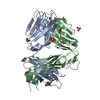
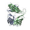
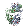
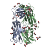
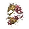
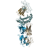
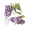
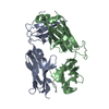
| Title | Crystal structure of a single-chain E/F type bilin lyase-isomerase MpeQ | ||||||
 Components Components | Bilin Lyase-Isomerase | ||||||
 Keywords Keywords | LYASE / bilin lyase-isomerase | ||||||
| Function / homology | E-Z type HEAT repeats / PBS lyase HEAT-like repeat / phycobilisome / HEAT repeats / Armadillo-like helical / Armadillo-type fold / lyase activity / Putative phycobilin:C-phycoerythrin II lyase Function and homology information Function and homology information | ||||||
| Biological species |  Synechococcus sp. P1 (bacteria) Synechococcus sp. P1 (bacteria) | ||||||
| Method |  X-RAY DIFFRACTION / SYNCHROTRON / SAD / Resolution: 2.5 Å X-RAY DIFFRACTION / SYNCHROTRON / SAD / Resolution: 2.5 Å | ||||||
 Authors Authors | Yang, X. / Kumarapperuma, I. | ||||||
| Funding support |  United States, 1items United States, 1items
| ||||||
 Citation Citation | Journal: Structure / Year: 2022 Title: Crystal structure and molecular mechanism of an E/F type bilin lyase-isomerase. Authors: Kumarapperuma, I. / Joseph, K.L. / Wang, C. / Biju, L.M. / Tom, I.P. / Weaver, K.D. / Grebert, T. / Partensky, F. / Schluchter, W.M. / Yang, X. | ||||||
| History |
|
- Structure visualization
| Structure viewer | Molecule: MolmilJmol/JSmol |
|---|
- Downloads & links
-Download
| PDBx/mmCIF format | 7mc4.cif.gz | 316.9 KB | Display | PDBx/mmCIF format |
|---|---|---|---|---|
| PDB format | pdb7mc4.ent.gz | 259.3 KB | Display | PDB format |
| PDBx/mmJSON format | 7mc4.json.gz | Tree view | PDBx/mmJSON format | |
| Others |  Other downloads Other downloads |
-Validation report
| Summary document | 7mc4_validation.pdf.gz | 436.8 KB | Display | wwPDB validaton report |
|---|---|---|---|---|
| Full document | 7mc4_full_validation.pdf.gz | 448.3 KB | Display | |
| Data in XML | 7mc4_validation.xml.gz | 31.4 KB | Display | |
| Data in CIF | 7mc4_validation.cif.gz | 44.3 KB | Display | |
| Arichive directory | https://data.pdbj.org/pub/pdb/validation_reports/mc/7mc4ftp://data.pdbj.org/pub/pdb/validation_reports/mc/7mc4 | HTTPS FTP |
-Related structure data


-Links
 PDBj
PDBj
- Assembly
| Deposited unit | 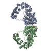
| ||||||||
|---|---|---|---|---|---|---|---|---|---|
| 1 | 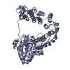
| ||||||||
| 2 | 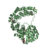
| ||||||||
| Unit cell |
| ||||||||
| Components on special symmetry positions |
|
-Components
| #1: Protein | Mass: 46678.965 Da / Num. of mol.: 2 Source method: isolated from a genetically manipulated source Source: (gene. exp.) Synechococcus sp. P1 (bacteria) / Strain: A15-62 / Gene: mpeY / Production host: #2: Water | ChemComp-HOH / |  Mass: 18.015 Da / Num. of mol.: 210 / Source method: isolated from a natural source / Formula: H2O Mass: 18.015 Da / Num. of mol.: 210 / Source method: isolated from a natural source / Formula: H2OHas ligand of interest | N | Has protein modification | Y | |
|---|
-Experimental details
-Experiment
| Experiment | Method: X-RAY DIFFRACTION / Number of used crystals: 1 |
|---|
- Sample preparation
| Crystal | Density Matthews: 3.63 Å3/Da / Density % sol: 66.11 % |
|---|---|
| Crystal grow | Temperature: 293 K / Method: vapor diffusion, hanging drop / pH: 7 Details: Protein concentration: 5 mg/mL, 2.1 M DL-Malic acid Temp details: room temperature |
-Data collection
| Diffraction | Mean temperature: 100 K / Serial crystal experiment: N |
|---|---|
| Diffraction source | Source: SYNCHROTRON / Site: APS / Beamline: 21-ID-F / Wavelength: 0.9787 Å |
| Detector | Type: MARMOSAIC 300 mm CCD / Detector: CCD / Date: Mar 3, 2020 |
| Radiation | Protocol: SINGLE WAVELENGTH / Monochromatic (M) / Laue (L): M / Scattering type: x-ray |
| Radiation wavelength | Wavelength: 0.9787 Å / Relative weight: 1 |
| Reflection | Resolution: 2.4→56 Å / Num. obs: 51398 / % possible obs: 99.78 % / Redundancy: 3.7 % / Biso Wilson estimate: 28.63 Å2 / CC1/2: 0.977 / Net I/σ(I): 3.4 |
| Reflection shell | Resolution: 2.4→2.44 Å / Num. unique obs: 2424 / CC1/2: 0.597 |
- Processing
| Software |
| |||||||||||||||||||||||||||||||||||||||||||||||||||||||||||||||||||||||||||||||||||||||||||||||||||||||||||||||||||||||||||||||||||||||||||||||||||||||||||||||||||||||||||||||
|---|---|---|---|---|---|---|---|---|---|---|---|---|---|---|---|---|---|---|---|---|---|---|---|---|---|---|---|---|---|---|---|---|---|---|---|---|---|---|---|---|---|---|---|---|---|---|---|---|---|---|---|---|---|---|---|---|---|---|---|---|---|---|---|---|---|---|---|---|---|---|---|---|---|---|---|---|---|---|---|---|---|---|---|---|---|---|---|---|---|---|---|---|---|---|---|---|---|---|---|---|---|---|---|---|---|---|---|---|---|---|---|---|---|---|---|---|---|---|---|---|---|---|---|---|---|---|---|---|---|---|---|---|---|---|---|---|---|---|---|---|---|---|---|---|---|---|---|---|---|---|---|---|---|---|---|---|---|---|---|---|---|---|---|---|---|---|---|---|---|---|---|---|---|---|---|---|
| Refinement | Method to determine structure: SAD / Resolution: 2.5→19.999 Å / SU ML: 0.35 / Cross valid method: THROUGHOUT / σ(F): 0 / Phase error: 32.43 / Stereochemistry target values: ML
| |||||||||||||||||||||||||||||||||||||||||||||||||||||||||||||||||||||||||||||||||||||||||||||||||||||||||||||||||||||||||||||||||||||||||||||||||||||||||||||||||||||||||||||||
| Solvent computation | Shrinkage radii: 0.9 Å / VDW probe radii: 1.11 Å / Solvent model: FLAT BULK SOLVENT MODEL | |||||||||||||||||||||||||||||||||||||||||||||||||||||||||||||||||||||||||||||||||||||||||||||||||||||||||||||||||||||||||||||||||||||||||||||||||||||||||||||||||||||||||||||||
| Displacement parameters | Biso max: 173.23 Å2 / Biso mean: 44.1166 Å2 / Biso min: 5.42 Å2 | |||||||||||||||||||||||||||||||||||||||||||||||||||||||||||||||||||||||||||||||||||||||||||||||||||||||||||||||||||||||||||||||||||||||||||||||||||||||||||||||||||||||||||||||
| Refinement step | Cycle: final / Resolution: 2.5→19.999 Å
| |||||||||||||||||||||||||||||||||||||||||||||||||||||||||||||||||||||||||||||||||||||||||||||||||||||||||||||||||||||||||||||||||||||||||||||||||||||||||||||||||||||||||||||||
| Refine LS restraints |
| |||||||||||||||||||||||||||||||||||||||||||||||||||||||||||||||||||||||||||||||||||||||||||||||||||||||||||||||||||||||||||||||||||||||||||||||||||||||||||||||||||||||||||||||
| LS refinement shell | Refine-ID: X-RAY DIFFRACTION / Rfactor Rfree error: 0
| |||||||||||||||||||||||||||||||||||||||||||||||||||||||||||||||||||||||||||||||||||||||||||||||||||||||||||||||||||||||||||||||||||||||||||||||||||||||||||||||||||||||||||||||
| Refinement TLS params. | Method: refined / Refine-ID: X-RAY DIFFRACTION
| |||||||||||||||||||||||||||||||||||||||||||||||||||||||||||||||||||||||||||||||||||||||||||||||||||||||||||||||||||||||||||||||||||||||||||||||||||||||||||||||||||||||||||||||
| Refinement TLS group |
|
