 Movie
Movie Controller


+ Open data

- Basic information
| Entry | Database: PDB / ID: 7mbf | ||||||
|---|---|---|---|---|---|---|---|
| Title | codeinone reductase isoform 1.3 Apo form | ||||||
 Components Components | NADPH-dependent codeinone reductase 1-3 | ||||||
 Keywords Keywords | OXIDOREDUCTASE / aldo-keto reductase / opium poppy / Benzylisoquinoline alkaloid / biosynthesis | ||||||
| Function / homology |  Function and homology information Function and homology informationcodeinone reductase (NADPH) / codeinone reductase (NADPH) activity / morphine metabolic process / codeine metabolic process / aldose reductase (NADPH) activity / oxidoreductase activity / cytosol Similarity search - Function | ||||||
| Biological species |  Papaver somniferum (opium poppy) Papaver somniferum (opium poppy) | ||||||
| Method |  X-RAY DIFFRACTION / SYNCHROTRON / MOLECULAR REPLACEMENT / Resolution: 2.4 Å X-RAY DIFFRACTION / SYNCHROTRON / MOLECULAR REPLACEMENT / Resolution: 2.4 Å | ||||||
 Authors Authors | Carr, S.C. / Ng, K.K.S. | ||||||
| Funding support |  Canada, 1items Canada, 1items
| ||||||
 Citation Citation | Journal: J.Biol.Chem. / Year: 2021 Title: Structural studies of codeinone reductase reveal novel insights into aldo-keto reductase function in benzylisoquinoline alkaloid biosynthesis. Authors: Carr, S.C. / Torres, M.A. / Morris, J.S. / Facchini, P.J. / Ng, K.K.S. | ||||||
| History |
|
- Structure visualization
| Structure viewer | Molecule: MolmilJmol/JSmol |
|---|
- Downloads & links
-Download
| PDBx/mmCIF format | 7mbf.cif.gz | 444.6 KB | Display | PDBx/mmCIF format |
|---|---|---|---|---|
| PDB format | pdb7mbf.ent.gz | 294.3 KB | Display | PDB format |
| PDBx/mmJSON format | 7mbf.json.gz | Tree view | PDBx/mmJSON format | |
| Others |  Other downloads Other downloads |
-Validation report
| Summary document | 7mbf_validation.pdf.gz | 481.8 KB | Display | wwPDB validaton report |
|---|---|---|---|---|
| Full document | 7mbf_full_validation.pdf.gz | 501.4 KB | Display | |
| Data in XML | 7mbf_validation.xml.gz | 63.1 KB | Display | |
| Data in CIF | 7mbf_validation.cif.gz | 85 KB | Display | |
| Arichive directory | https://data.pdbj.org/pub/pdb/validation_reports/mb/7mbfftp://data.pdbj.org/pub/pdb/validation_reports/mb/7mbf | HTTPS FTP |
-Related structure data
| Related structure data | 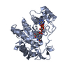 1zgdS S: Starting model for refinement |
|---|---|
| Similar structure data |


-Links
 PDBj
PDBj
- Assembly
| Deposited unit | 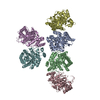
| ||||||||||||
|---|---|---|---|---|---|---|---|---|---|---|---|---|---|
| 1 | 
| ||||||||||||
| 2 | 
| ||||||||||||
| 3 | 
| ||||||||||||
| 4 | 
| ||||||||||||
| 5 | 
| ||||||||||||
| 6 | 
| ||||||||||||
| Unit cell |
|
-Components
| #1: Protein | Mass: 36341.902 Da / Num. of mol.: 6 Source method: isolated from a genetically manipulated source Source: (gene. exp.) Papaver somniferum (opium poppy) / Gene: COR1.3 / Plasmid: pET47b / Production host:  #2: Water | ChemComp-HOH / |  Mass: 18.015 Da / Num. of mol.: 164 / Source method: isolated from a natural source / Formula: H2O Mass: 18.015 Da / Num. of mol.: 164 / Source method: isolated from a natural source / Formula: H2OHas protein modification | Y | |
|---|
-Experimental details
-Experiment
| Experiment | Method: X-RAY DIFFRACTION / Number of used crystals: 1 |
|---|
- Sample preparation
| Crystal | Density Matthews: 2.39 Å3/Da / Density % sol: 48.7 % / Description: prism |
|---|---|
| Crystal grow | Temperature: 298 K / Method: vapor diffusion, hanging drop / pH: 8 Details: 24% polyethylene glycol 3350, 0.35M sodium chloride, 8% glycerol, 2mM DTT, 0.1M Tris-HCl pH 8.0 |
-Data collection
| Diffraction | Mean temperature: 100 K / Serial crystal experiment: N |
|---|---|
| Diffraction source | Source: SYNCHROTRON / Site: SSRL  / Beamline: BL12-2 / Wavelength: 0.97946 Å / Beamline: BL12-2 / Wavelength: 0.97946 Å |
| Detector | Type: DECTRIS PILATUS 6M / Detector: PIXEL / Date: Jul 4, 2019 Details: Flat Si Rh coated M0, Kirkpatrick-Baez flat bent Si M1 & M2 |
| Radiation | Monochromator: Liquid nitrogen-cooled double crystal Si(111) Protocol: SINGLE WAVELENGTH / Monochromatic (M) / Laue (L): M / Scattering type: x-ray |
| Radiation wavelength | Wavelength: 0.97946 Å / Relative weight: 1 |
| Reflection | Resolution: 2.4→50 Å / Num. obs: 68275 / % possible obs: 86.2 % / Observed criterion σ(I): -3 / Redundancy: 2.9 % / Biso Wilson estimate: 39.01 Å2 / CC1/2: 0.966 / CC star: 0.991 / Rmerge(I) obs: 0.214 / Rpim(I) all: 0.143 / Rrim(I) all: 0.258 / Rsym value: 0.214 / Net I/av σ(I): 3.88 / Net I/σ(I): 2.8 |
| Reflection shell | Resolution: 2.4→2.49 Å / Redundancy: 1.7 % / Rmerge(I) obs: 0.958 / Mean I/σ(I) obs: 0.58 / Num. unique obs: 3743 / CC1/2: 0.523 / CC star: 0.827 / Rpim(I) all: 0.735 / Rrim(I) all: 1.215 / Rsym value: 0.958 / % possible all: 47.6 |
- Processing
| Software |
| |||||||||||||||||||||||||||||||||||||||||||||||||||||||||||||||||||||||||||||||||||||||||||||||||||||||||
|---|---|---|---|---|---|---|---|---|---|---|---|---|---|---|---|---|---|---|---|---|---|---|---|---|---|---|---|---|---|---|---|---|---|---|---|---|---|---|---|---|---|---|---|---|---|---|---|---|---|---|---|---|---|---|---|---|---|---|---|---|---|---|---|---|---|---|---|---|---|---|---|---|---|---|---|---|---|---|---|---|---|---|---|---|---|---|---|---|---|---|---|---|---|---|---|---|---|---|---|---|---|---|---|---|---|---|
| Refinement | Method to determine structure: MOLECULAR REPLACEMENT Starting model: 1zgd Resolution: 2.4→43.37 Å / SU ML: 0.4144 / Cross valid method: FREE R-VALUE / Phase error: 32.3644 Stereochemistry target values: GeoStd + Monomer Library + CDL v1.2
| |||||||||||||||||||||||||||||||||||||||||||||||||||||||||||||||||||||||||||||||||||||||||||||||||||||||||
| Solvent computation | Shrinkage radii: 0.9 Å / VDW probe radii: 1.11 Å / Solvent model: FLAT BULK SOLVENT MODEL | |||||||||||||||||||||||||||||||||||||||||||||||||||||||||||||||||||||||||||||||||||||||||||||||||||||||||
| Displacement parameters | Biso mean: 50.2 Å2 | |||||||||||||||||||||||||||||||||||||||||||||||||||||||||||||||||||||||||||||||||||||||||||||||||||||||||
| Refinement step | Cycle: LAST / Resolution: 2.4→43.37 Å
| |||||||||||||||||||||||||||||||||||||||||||||||||||||||||||||||||||||||||||||||||||||||||||||||||||||||||
| Refine LS restraints |
| |||||||||||||||||||||||||||||||||||||||||||||||||||||||||||||||||||||||||||||||||||||||||||||||||||||||||
| LS refinement shell |
|
