 Movie
Movie Controller

[English] 日本語
 Yorodumi
- PDB-7lzy: Structure of SARS-CoV-2 3CL protease in complex with inhibitor 3c -
+ Open data

- Basic information
| Entry | Database: PDB / ID: 7lzy | ||||||
|---|---|---|---|---|---|---|---|
| Title | Structure of SARS-CoV-2 3CL protease in complex with inhibitor 3c | ||||||
 Components Components | 3C-like proteinase | ||||||
 Keywords Keywords | HYDROLASE/INHIBITOR / COVID-19 / PROTEASE / severe acute respiratory syndrome coronavirus 2 / SARS-CoV-2 3CL protease Inhhibitors / HYDROLASE-INHIBITOR complex | ||||||
| Function / homology |  Function and homology information Function and homology informationprotein guanylyltransferase activity / RNA endonuclease activity producing 3'-phosphomonoesters, hydrolytic mechanism / mRNA guanylyltransferase activity / 5'-3' RNA helicase activity / Lyases; Phosphorus-oxygen lyases / Assembly of the SARS-CoV-2 Replication-Transcription Complex (RTC) / symbiont-mediated suppression of host cytoplasmic pattern recognition receptor signaling pathway via inhibition of TBK1 activity / Maturation of replicase proteins / ISG15-specific peptidase activity / TRAF3-dependent IRF activation pathway ...protein guanylyltransferase activity / RNA endonuclease activity producing 3'-phosphomonoesters, hydrolytic mechanism / mRNA guanylyltransferase activity / 5'-3' RNA helicase activity / Lyases; Phosphorus-oxygen lyases / Assembly of the SARS-CoV-2 Replication-Transcription Complex (RTC) / symbiont-mediated suppression of host cytoplasmic pattern recognition receptor signaling pathway via inhibition of TBK1 activity / Maturation of replicase proteins / ISG15-specific peptidase activity / TRAF3-dependent IRF activation pathway / Transcription of SARS-CoV-2 sgRNAs / Translation of Replicase and Assembly of the Replication Transcription Complex / snRNP Assembly / Replication of the SARS-CoV-2 genome / Hydrolases; Acting on ester bonds; Exoribonucleases producing 5'-phosphomonoesters / double membrane vesicle viral factory outer membrane / host cell endoplasmic reticulum-Golgi intermediate compartment / SARS coronavirus main proteinase / 5'-3' DNA helicase activity / 3'-5'-RNA exonuclease activity / host cell endosome / symbiont-mediated degradation of host mRNA / mRNA guanylyltransferase / symbiont-mediated suppression of host ISG15-protein conjugation / G-quadruplex RNA binding / symbiont-mediated suppression of host toll-like receptor signaling pathway / symbiont-mediated suppression of host cytoplasmic pattern recognition receptor signaling pathway via inhibition of IRF3 activity / omega peptidase activity / mRNA (guanine-N7)-methyltransferase / SARS-CoV-2 modulates host translation machinery / methyltransferase cap1 / host cell Golgi apparatus / symbiont-mediated suppression of host NF-kappaB cascade / symbiont-mediated perturbation of host ubiquitin-like protein modification / DNA helicase / methyltransferase cap1 activity / ubiquitinyl hydrolase 1 / cysteine-type deubiquitinase activity / mRNA 5'-cap (guanine-N7-)-methyltransferase activity / Hydrolases; Acting on peptide bonds (peptidases); Cysteine endopeptidases / single-stranded RNA binding / host cell perinuclear region of cytoplasm / regulation of autophagy / viral protein processing / lyase activity / host cell endoplasmic reticulum membrane / RNA helicase / symbiont-mediated suppression of host type I interferon-mediated signaling pathway / symbiont-mediated suppression of host gene expression / copper ion binding / viral translational frameshifting / symbiont-mediated activation of host autophagy / RNA-directed RNA polymerase / cysteine-type endopeptidase activity / viral RNA genome replication / RNA-directed RNA polymerase activity / lipid binding / DNA-templated transcription / SARS-CoV-2 activates/modulates innate and adaptive immune responses / host cell nucleus / ATP hydrolysis activity / proteolysis / RNA binding / zinc ion binding / ATP binding / membrane Similarity search - Function | ||||||
| Biological species |   Severe acute respiratory syndrome coronavirus 2 Severe acute respiratory syndrome coronavirus 2 | ||||||
| Method |  X-RAY DIFFRACTION / SYNCHROTRON / MOLECULAR REPLACEMENT / Resolution: 1.85 Å X-RAY DIFFRACTION / SYNCHROTRON / MOLECULAR REPLACEMENT / Resolution: 1.85 Å | ||||||
 Authors Authors | Lovell, S. / Kashipathy, M.M. / Battaile, K.P. / Chamandi, S.D. / Rathnayake, A.D. / Kim, Y. / Perera, K.D. / Jesri, A.R.M. / Nguyen, H.N. / Baird, M.A. ...Lovell, S. / Kashipathy, M.M. / Battaile, K.P. / Chamandi, S.D. / Rathnayake, A.D. / Kim, Y. / Perera, K.D. / Jesri, A.R.M. / Nguyen, H.N. / Baird, M.A. / Miller, M.J. / Groutas, W.C. / Chang, K.O. | ||||||
| Funding support |  United States, 1items United States, 1items
| ||||||
 Citation Citation | Journal: J.Med.Chem. / Year: 2021 Title: Structure-Guided Design of Potent Inhibitors of SARS-CoV-2 3CL Protease: Structural, Biochemical, and Cell-Based Studies. Authors: Dampalla, C.S. / Rathnayake, A.D. / Perera, K.D. / Jesri, A.M. / Nguyen, H.N. / Miller, M.J. / Thurman, H.A. / Zheng, J. / Kashipathy, M.M. / Battaile, K.P. / Lovell, S. / Perlman, S. / Kim, ...Authors: Dampalla, C.S. / Rathnayake, A.D. / Perera, K.D. / Jesri, A.M. / Nguyen, H.N. / Miller, M.J. / Thurman, H.A. / Zheng, J. / Kashipathy, M.M. / Battaile, K.P. / Lovell, S. / Perlman, S. / Kim, Y. / Groutas, W.C. / Chang, K.O. | ||||||
| History |
|
- Structure visualization
| Structure viewer | Molecule: MolmilJmol/JSmol |
|---|
- Downloads & links
-Download
| PDBx/mmCIF format | 7lzy.cif.gz | 76.9 KB | Display | PDBx/mmCIF format |
|---|---|---|---|---|
| PDB format | pdb7lzy.ent.gz | 54.2 KB | Display | PDB format |
| PDBx/mmJSON format | 7lzy.json.gz | Tree view | PDBx/mmJSON format | |
| Others |  Other downloads Other downloads |
-Validation report
| Summary document | 7lzy_validation.pdf.gz | 455.2 KB | Display | wwPDB validaton report |
|---|---|---|---|---|
| Full document | 7lzy_full_validation.pdf.gz | 455.3 KB | Display | |
| Data in XML | 7lzy_validation.xml.gz | 15.7 KB | Display | |
| Data in CIF | 7lzy_validation.cif.gz | 20.9 KB | Display | |
| Arichive directory | https://data.pdbj.org/pub/pdb/validation_reports/lz/7lzyftp://data.pdbj.org/pub/pdb/validation_reports/lz/7lzy | HTTPS FTP |
-Related structure data
| Related structure data |  7lztC  7lzuC  7lzvC  7lzwC  7lzxC  7lzzC  7m00C  7m01C 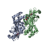 7m02C 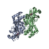 7m03C 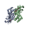 7m04C 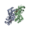 6xmkS S: Starting model for refinement C: citing same article ( |
|---|---|
| Similar structure data |


-Links
 PDBj
PDBj


- Assembly
| Deposited unit | 
| |||||||||
|---|---|---|---|---|---|---|---|---|---|---|
| 1 | 
| |||||||||
| Unit cell |
| |||||||||
| Components on special symmetry positions |
|
-Components
| #1: Protein | Mass: 34068.805 Da / Num. of mol.: 1 Source method: isolated from a genetically manipulated source Source: (gene. exp.) Severe acute respiratory syndrome coronavirus 2Gene: rep, 1a-1b / Plasmid: pET28 / Production host:  References: UniProt: P0DTD1, SARS coronavirus main proteinase |
|---|---|
| #2: Chemical | ChemComp-YMJ / (  Mass: 519.652 Da / Num. of mol.: 1 / Source method: obtained synthetically / Formula: C23H41N3O8S Mass: 519.652 Da / Num. of mol.: 1 / Source method: obtained synthetically / Formula: C23H41N3O8S |
| #3: Chemical | ChemComp-YMM / (  Mass: 519.652 Da / Num. of mol.: 1 / Source method: isolated from a natural source / Formula: C23H41N3O8S Mass: 519.652 Da / Num. of mol.: 1 / Source method: isolated from a natural source / Formula: C23H41N3O8S |
| #4: Water | ChemComp-HOH /  Mass: 18.015 Da / Num. of mol.: 137 / Source method: isolated from a natural source / Formula: H2O Mass: 18.015 Da / Num. of mol.: 137 / Source method: isolated from a natural source / Formula: H2O |
| Has ligand of interest | Y |
| Has protein modification | Y |
-Experimental details
-Experiment
| Experiment | Method: X-RAY DIFFRACTION / Number of used crystals: 1 |
|---|
- Sample preparation
| Crystal | Density Matthews: 2 Å3/Da / Density % sol: 38.58 % |
|---|---|
| Crystal grow | Temperature: 293 K / Method: vapor diffusion, sitting drop / pH: 5.5 Details: 17% w/v PEG10000, 100 mM Bis-Tris, 100 mM ammonium acetate |
-Data collection
| Diffraction | Mean temperature: 100 K / Serial crystal experiment: N | ||||||||||||||||||||||||
|---|---|---|---|---|---|---|---|---|---|---|---|---|---|---|---|---|---|---|---|---|---|---|---|---|---|
| Diffraction source | Source: SYNCHROTRON / Site: APS / Beamline: 17-ID / Wavelength: 1 Å | ||||||||||||||||||||||||
| Detector | Type: DECTRIS PILATUS 6M / Detector: PIXEL / Date: Feb 14, 2021 | ||||||||||||||||||||||||
| Radiation | Protocol: SINGLE WAVELENGTH / Monochromatic (M) / Laue (L): M / Scattering type: x-ray | ||||||||||||||||||||||||
| Radiation wavelength | Wavelength: 1 Å / Relative weight: 1 | ||||||||||||||||||||||||
| Reflection | Resolution: 1.85→48.03 Å / Num. obs: 23131 / % possible obs: 100 % / Redundancy: 6.9 % / Biso Wilson estimate: 30.85 Å2 / CC1/2: 0.998 / Rmerge(I) obs: 0.085 / Net I/σ(I): 11.4 / Num. measured all: 158821 | ||||||||||||||||||||||||
| Reflection shell | Diffraction-ID: 1
|
- Processing
| Software |
| ||||||||||||||||||||||||||||||||||||||||||||||||||||||
|---|---|---|---|---|---|---|---|---|---|---|---|---|---|---|---|---|---|---|---|---|---|---|---|---|---|---|---|---|---|---|---|---|---|---|---|---|---|---|---|---|---|---|---|---|---|---|---|---|---|---|---|---|---|---|---|
| Refinement | Method to determine structure: MOLECULAR REPLACEMENT Starting model: PDB entry 6XMK Resolution: 1.85→39.39 Å / SU ML: 0.22 / Cross valid method: THROUGHOUT / σ(F): 1.01 / Phase error: 23.72 / Stereochemistry target values: ML
| ||||||||||||||||||||||||||||||||||||||||||||||||||||||
| Solvent computation | Shrinkage radii: 0.9 Å / VDW probe radii: 1.11 Å / Solvent model: FLAT BULK SOLVENT MODEL | ||||||||||||||||||||||||||||||||||||||||||||||||||||||
| Displacement parameters | Biso max: 81.6 Å2 / Biso mean: 35.6265 Å2 / Biso min: 16.03 Å2 | ||||||||||||||||||||||||||||||||||||||||||||||||||||||
| Refinement step | Cycle: final / Resolution: 1.85→39.39 Å
| ||||||||||||||||||||||||||||||||||||||||||||||||||||||
| LS refinement shell | Refine-ID: X-RAY DIFFRACTION / Rfactor Rfree error: 0 / Total num. of bins used: 8 / % reflection obs: 100 %
|
