 Movie
Movie Controller

[English] 日本語
 Yorodumi
- PDB-7lt9: Crystal structure of Ras suppressor-1 in complex with PINCH-1 LIM... -
+ Open data

- Basic information
| Entry | Database: PDB / ID: 7lt9 | ||||||
|---|---|---|---|---|---|---|---|
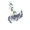
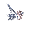
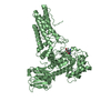
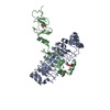
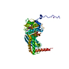
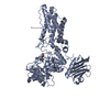
| Title | Crystal structure of Ras suppressor-1 in complex with PINCH-1 LIM4-5 domains | ||||||
 Components Components |
| ||||||
 Keywords Keywords | CELL ADHESION / leucine-rich repeat LIM domain | ||||||
| Function / homology |  Function and homology information Function and homology informationRegulation of cytoskeletal remodeling and cell spreading by IPP complex components / positive regulation of integrin-mediated signaling pathway / cell-cell junction organization / Cell-extracellular matrix interactions / cellular response to transforming growth factor beta stimulus / positive regulation of substrate adhesion-dependent cell spreading / cell-matrix adhesion / tumor necrosis factor-mediated signaling pathway / cell-cell adhesion / cell-cell junction ...Regulation of cytoskeletal remodeling and cell spreading by IPP complex components / positive regulation of integrin-mediated signaling pathway / cell-cell junction organization / Cell-extracellular matrix interactions / cellular response to transforming growth factor beta stimulus / positive regulation of substrate adhesion-dependent cell spreading / cell-matrix adhesion / tumor necrosis factor-mediated signaling pathway / cell-cell adhesion / cell-cell junction / actin binding / Ras protein signal transduction / positive regulation of canonical NF-kappaB signal transduction / focal adhesion / negative regulation of DNA-templated transcription / protein kinase binding / perinuclear region of cytoplasm / signal transduction / extracellular exosome / zinc ion binding / plasma membrane / cytosol / cytoplasm Similarity search - Function | ||||||
| Biological species |  Homo sapiens (human) Homo sapiens (human) | ||||||
| Method |  X-RAY DIFFRACTION / SYNCHROTRON / MOLECULAR REPLACEMENT / molecular replacement / Resolution: 3.05011197028 Å X-RAY DIFFRACTION / SYNCHROTRON / MOLECULAR REPLACEMENT / molecular replacement / Resolution: 3.05011197028 Å | ||||||
 Authors Authors | Fukuda, K. / Qin, J. | ||||||
| Funding support |  United States, 1items United States, 1items
| ||||||
 Citation Citation | Journal: J.Biol.Chem. / Year: 2021 Title: Molecular basis for Ras suppressor-1 binding to PINCH-1 in focal adhesion assembly. Authors: Fukuda, K. / Lu, F. / Qin, J. | ||||||
| History |
|
- Structure visualization
| Structure viewer | Molecule: MolmilJmol/JSmol |
|---|
- Downloads & links
-Download
| PDBx/mmCIF format | 7lt9.cif.gz | 110.2 KB | Display | PDBx/mmCIF format |
|---|---|---|---|---|
| PDB format | pdb7lt9.ent.gz | 67.6 KB | Display | PDB format |
| PDBx/mmJSON format | 7lt9.json.gz | Tree view | PDBx/mmJSON format | |
| Others |  Other downloads Other downloads |
-Validation report
| Arichive directory | https://data.pdbj.org/pub/pdb/validation_reports/lt/7lt9ftp://data.pdbj.org/pub/pdb/validation_reports/lt/7lt9 | HTTPS FTP |
|---|
-Related structure data
| Related structure data |  7lt8SC S: Starting model for refinement C: citing same article ( |
|---|---|
| Similar structure data |


-Links
 PDBj
PDBj


- Assembly
| Deposited unit | 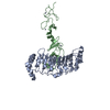
| ||||||||||
|---|---|---|---|---|---|---|---|---|---|---|---|
| 1 |
| ||||||||||
| Unit cell |
|
-Components
| #1: Protein | Mass: 31861.537 Da / Num. of mol.: 1 Source method: isolated from a genetically manipulated source Source: (gene. exp.) Homo sapiens (human) / Gene: RSU1, RSP1 / Production host:   Spodoptera frugiperda (fall armyworm) / Strain (production host): Sf-9 / References: UniProt: Q15404 Spodoptera frugiperda (fall armyworm) / Strain (production host): Sf-9 / References: UniProt: Q15404 | ||
|---|---|---|---|
| #2: Protein | Mass: 16189.122 Da / Num. of mol.: 1 / Fragment: LIM domain (UNP residues 193-329) Source method: isolated from a genetically manipulated source Source: (gene. exp.) Homo sapiens (human) / Gene: LIMS1, PINCH, PINCH1 / Plasmid: pGEX4T1 / Production host:  | ||
| #3: Chemical | ChemComp-ZN /   Mass: 65.409 Da / Num. of mol.: 4 / Source method: obtained synthetically / Formula: Zn / Feature type: SUBJECT OF INVESTIGATION Mass: 65.409 Da / Num. of mol.: 4 / Source method: obtained synthetically / Formula: Zn / Feature type: SUBJECT OF INVESTIGATIONHas ligand of interest | Y | |
-Experimental details
-Experiment
| Experiment | Method: X-RAY DIFFRACTION / Number of used crystals: 1 |
|---|
- Sample preparation
| Crystal | Density Matthews: 3.66 Å3/Da / Density % sol: 66.42 % |
|---|---|
| Crystal grow | Temperature: 296 K / Method: vapor diffusion / pH: 7.5 / Details: PEG8000 |
-Data collection
| Diffraction | Mean temperature: 100 K / Serial crystal experiment: N |
|---|---|
| Diffraction source | Source: SYNCHROTRON / Site: APS / Beamline: 19-BM / Wavelength: 0.97919 Å |
| Detector | Type: ADSC QUANTUM 210r / Detector: CCD / Date: Mar 29, 2019 |
| Radiation | Monochromator: double crystal Si(111) / Protocol: SINGLE WAVELENGTH / Monochromatic (M) / Laue (L): M / Scattering type: x-ray |
| Radiation wavelength | Wavelength: 0.97919 Å / Relative weight: 1 |
| Reflection | Resolution: 3.05011197028→50 Å / Num. obs: 13446 / % possible obs: 97.1 % / Redundancy: 4.6 % / Biso Wilson estimate: 67.4053598249 Å2 / CC1/2: 0.992 / CC star: 0.998 / Rmerge(I) obs: 0.091 / Net I/σ(I): 27.76225 |
| Reflection shell | Resolution: 3.05011197028→3.1 Å / Rmerge(I) obs: 0.852 / Num. unique obs: 669 / CC1/2: 0.731 / CC star: 0.919 / % possible all: 99.7 |
-Phasing
| Phasing | Method: molecular replacement |
|---|
- Processing
| Software |
| |||||||||||||||||||||||||||||||||||
|---|---|---|---|---|---|---|---|---|---|---|---|---|---|---|---|---|---|---|---|---|---|---|---|---|---|---|---|---|---|---|---|---|---|---|---|---|
| Refinement | Method to determine structure: MOLECULAR REPLACEMENT Starting model: PDB entry 7LT8 Resolution: 3.05011197028→34.5707614021 Å / SU ML: 0.399321051542 / Cross valid method: THROUGHOUT / σ(F): 1.33638551595 / Phase error: 23.6012965098 Stereochemistry target values: GeoStd + Monomer Library + CDL v1.2
| |||||||||||||||||||||||||||||||||||
| Solvent computation | Shrinkage radii: 0.9 Å / VDW probe radii: 1.11 Å / Solvent model: FLAT BULK SOLVENT MODEL | |||||||||||||||||||||||||||||||||||
| Displacement parameters | Biso mean: 77.314907178 Å2 | |||||||||||||||||||||||||||||||||||
| Refinement step | Cycle: LAST / Resolution: 3.05011197028→34.5707614021 Å
| |||||||||||||||||||||||||||||||||||
| Refine LS restraints |
| |||||||||||||||||||||||||||||||||||
| LS refinement shell |
|
