 Movie
Movie Controller

[English] 日本語
 Yorodumi
- PDB-7lr9: Crystal structure of KPC-2 S70G/T215P mutant with hydrolyzed imipenem -
+ Open data

- Basic information
| Entry | Database: PDB / ID: 7lr9 | ||||||
|---|---|---|---|---|---|---|---|
| Title | Crystal structure of KPC-2 S70G/T215P mutant with hydrolyzed imipenem | ||||||
 Components Components | Carbapenem-hydrolyzing beta-lactamase KPC | ||||||
 Keywords Keywords | HYDROLASE/ANTIBIOTIC / KPC / Carbapenemase / Beta-lactamase / Hydrolase / Antibiotic Resistance / Enzyme / Beta-lactam / antibiotics / HYDROLASE-ANTIBIOTIC complex | ||||||
| Function / homology |  Function and homology information Function and homology informationAntimicrobial resistance / beta-lactam antibiotic catabolic process / beta-lactamase activity / beta-lactamase / response to antibiotic / cytosol Similarity search - Function | ||||||
| Biological species |  Klebsiella pneumoniae (bacteria) Klebsiella pneumoniae (bacteria) | ||||||
| Method |  X-RAY DIFFRACTION / SYNCHROTRON / MOLECULAR REPLACEMENT / Resolution: 1.47 Å X-RAY DIFFRACTION / SYNCHROTRON / MOLECULAR REPLACEMENT / Resolution: 1.47 Å | ||||||
 Authors Authors | Furey, I. / Palzkill, T. / Hu, L. / Prasad, B.V.V. | ||||||
| Funding support |  United States, 1items United States, 1items
| ||||||
 Citation Citation | Journal: To Be Published Title: Crystal structure of KPC-2 T215P mutant Authors: Furey, I.M. / Palzkill, T. | ||||||
| History |
|
- Structure visualization
| Structure viewer | Molecule: MolmilJmol/JSmol |
|---|
- Downloads & links
-Download
| PDBx/mmCIF format | 7lr9.cif.gz | 136 KB | Display | PDBx/mmCIF format |
|---|---|---|---|---|
| PDB format | pdb7lr9.ent.gz | 94.2 KB | Display | PDB format |
| PDBx/mmJSON format | 7lr9.json.gz | Tree view | PDBx/mmJSON format | |
| Others |  Other downloads Other downloads |
-Validation report
| Arichive directory | https://data.pdbj.org/pub/pdb/validation_reports/lr/7lr9ftp://data.pdbj.org/pub/pdb/validation_reports/lr/7lr9 | HTTPS FTP |
|---|
-Related structure data
| Related structure data | 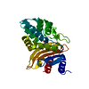 3c5aS S: Starting model for refinement |
|---|---|
| Similar structure data |
-Links
 PDBj
PDBj


- Assembly
| Deposited unit | 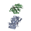
| ||||||||||||
|---|---|---|---|---|---|---|---|---|---|---|---|---|---|
| 1 | 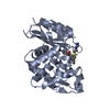
| ||||||||||||
| 2 | 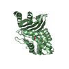
| ||||||||||||
| Unit cell |
|
-Components
| #1: Protein | Mass: 28314.910 Da / Num. of mol.: 2 / Mutation: S70G, T215P Source method: isolated from a genetically manipulated source Source: (gene. exp.) Klebsiella pneumoniae (bacteria) / Gene: bla, kpc, kpc1 / Production host: #2: Chemical | 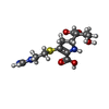  Mass: 317.361 Da / Num. of mol.: 2 / Source method: obtained synthetically / Formula: C12H19N3O5S / Feature type: SUBJECT OF INVESTIGATION Mass: 317.361 Da / Num. of mol.: 2 / Source method: obtained synthetically / Formula: C12H19N3O5S / Feature type: SUBJECT OF INVESTIGATION#3: Water | ChemComp-HOH / |  Mass: 18.015 Da / Num. of mol.: 595 / Source method: isolated from a natural source / Formula: H2O Mass: 18.015 Da / Num. of mol.: 595 / Source method: isolated from a natural source / Formula: H2OHas ligand of interest | Y | Has protein modification | Y | |
|---|
-Experimental details
-Experiment
| Experiment | Method: X-RAY DIFFRACTION / Number of used crystals: 1 |
|---|
- Sample preparation
| Crystal | Density Matthews: 1.91 Å3/Da / Density % sol: 35.47 % |
|---|---|
| Crystal grow | Temperature: 298 K / Method: vapor diffusion, hanging drop Details: 25% PEG 8,000, 0.1M KsCN, 0.1M Sodium Acetate pH:4.5 |
-Data collection
| Diffraction | Mean temperature: 100 K / Serial crystal experiment: N |
|---|---|
| Diffraction source | Source: SYNCHROTRON / Site: ALS / Beamline: 8.2.1 / Wavelength: 0.999995 Å |
| Detector | Type: ADSC QUANTUM 315r / Detector: CCD / Date: Mar 13, 2019 |
| Radiation | Protocol: SINGLE WAVELENGTH / Monochromatic (M) / Laue (L): M / Scattering type: x-ray |
| Radiation wavelength | Wavelength: 0.999995 Å / Relative weight: 1 |
| Reflection | Resolution: 1.47→37.64 Å / Num. obs: 68235 / % possible obs: 95.86 % / Redundancy: 3 % / Biso Wilson estimate: 10.92 Å2 / CC1/2: 0.997 / Rmerge(I) obs: 0.04879 / Net I/σ(I): 12.4 |
| Reflection shell | Resolution: 1.47→1.523 Å / Rmerge(I) obs: 0.101 / Num. unique obs: 6730 / CC1/2: 0.984 |
- Processing
| Software |
| |||||||||||||||||||||||||||||||||||||||||||||||||||||||||||||||||||||||||||||||||||||||||||||||||||||||||||||||||||||||||||||||||||||||||||||||||||||||||||||||||||||||||||||||
|---|---|---|---|---|---|---|---|---|---|---|---|---|---|---|---|---|---|---|---|---|---|---|---|---|---|---|---|---|---|---|---|---|---|---|---|---|---|---|---|---|---|---|---|---|---|---|---|---|---|---|---|---|---|---|---|---|---|---|---|---|---|---|---|---|---|---|---|---|---|---|---|---|---|---|---|---|---|---|---|---|---|---|---|---|---|---|---|---|---|---|---|---|---|---|---|---|---|---|---|---|---|---|---|---|---|---|---|---|---|---|---|---|---|---|---|---|---|---|---|---|---|---|---|---|---|---|---|---|---|---|---|---|---|---|---|---|---|---|---|---|---|---|---|---|---|---|---|---|---|---|---|---|---|---|---|---|---|---|---|---|---|---|---|---|---|---|---|---|---|---|---|---|---|---|---|---|
| Refinement | Method to determine structure: MOLECULAR REPLACEMENT Starting model: 3C5A Resolution: 1.47→37.64 Å / SU ML: 0.1218 / Cross valid method: FREE R-VALUE / σ(F): 2.01 / Phase error: 17.6412 / Stereochemistry target values: CDL v1.2
| |||||||||||||||||||||||||||||||||||||||||||||||||||||||||||||||||||||||||||||||||||||||||||||||||||||||||||||||||||||||||||||||||||||||||||||||||||||||||||||||||||||||||||||||
| Solvent computation | Shrinkage radii: 0.9 Å / VDW probe radii: 1.11 Å / Solvent model: FLAT BULK SOLVENT MODEL | |||||||||||||||||||||||||||||||||||||||||||||||||||||||||||||||||||||||||||||||||||||||||||||||||||||||||||||||||||||||||||||||||||||||||||||||||||||||||||||||||||||||||||||||
| Displacement parameters | Biso mean: 15.15 Å2 | |||||||||||||||||||||||||||||||||||||||||||||||||||||||||||||||||||||||||||||||||||||||||||||||||||||||||||||||||||||||||||||||||||||||||||||||||||||||||||||||||||||||||||||||
| Refinement step | Cycle: LAST / Resolution: 1.47→37.64 Å
| |||||||||||||||||||||||||||||||||||||||||||||||||||||||||||||||||||||||||||||||||||||||||||||||||||||||||||||||||||||||||||||||||||||||||||||||||||||||||||||||||||||||||||||||
| Refine LS restraints |
| |||||||||||||||||||||||||||||||||||||||||||||||||||||||||||||||||||||||||||||||||||||||||||||||||||||||||||||||||||||||||||||||||||||||||||||||||||||||||||||||||||||||||||||||
| LS refinement shell |
|
