 Movie
Movie Controller

[English] 日本語
 Yorodumi
- PDB-7lpq: Crystal structure of Cryptococcus neoformans sterylglucosidase 1 ... -
+ Open data

- Basic information
| Entry | Database: PDB / ID: 7lpq | |||||||||
|---|---|---|---|---|---|---|---|---|---|---|
| Title | Crystal structure of Cryptococcus neoformans sterylglucosidase 1 with hit 9 | |||||||||
 Components Components | Cytoplasmic protein | |||||||||
 Keywords Keywords | HYDROLASE / glucosidase / sterylglucosidase | |||||||||
| Function / homology |  Function and homology information Function and homology informationergosteryl 3-beta-D-glucoside catabolic process / steryl-beta-glucosidase activity / polysaccharide catabolic process Similarity search - Function | |||||||||
| Biological species |  Cryptococcus neoformans var. grubii serotype A (fungus) Cryptococcus neoformans var. grubii serotype A (fungus) | |||||||||
| Method |  X-RAY DIFFRACTION / SYNCHROTRON / MOLECULAR REPLACEMENT / Resolution: 2.32 Å X-RAY DIFFRACTION / SYNCHROTRON / MOLECULAR REPLACEMENT / Resolution: 2.32 Å | |||||||||
 Authors Authors | Pereira de Sa, N. / Del Poeta, M. / Airola, M.V. | |||||||||
| Funding support |  United States, 2items United States, 2items
| |||||||||
 Citation Citation | Journal: Nat Commun / Year: 2021 Title: Structure and inhibition of Cryptococcus neoformans sterylglucosidase to develop antifungal agents. Authors: Pereira de Sa, N. / Taouil, A. / Kim, J. / Clement, T. / Hoffmann, R.M. / Burke, J.E. / Rizzo, R.C. / Ojima, I. / Del Poeta, M. / Airola, M.V. | |||||||||
| History |
|
- Structure visualization
| Structure viewer | Molecule: MolmilJmol/JSmol |
|---|
- Downloads & links
-Download
| PDBx/mmCIF format | 7lpq.cif.gz | 1.6 MB | Display | PDBx/mmCIF format |
|---|---|---|---|---|
| PDB format | pdb7lpq.ent.gz | 1.4 MB | Display | PDB format |
| PDBx/mmJSON format | 7lpq.json.gz | Tree view | PDBx/mmJSON format | |
| Others |  Other downloads Other downloads |
-Validation report
| Summary document | 7lpq_validation.pdf.gz | 1.4 MB | Display | wwPDB validaton report |
|---|---|---|---|---|
| Full document | 7lpq_full_validation.pdf.gz | 1.5 MB | Display | |
| Data in XML | 7lpq_validation.xml.gz | 116 KB | Display | |
| Data in CIF | 7lpq_validation.cif.gz | 168.6 KB | Display | |
| Arichive directory | https://data.pdbj.org/pub/pdb/validation_reports/lp/7lpqftp://data.pdbj.org/pub/pdb/validation_reports/lp/7lpq | HTTPS FTP |
-Related structure data
| Related structure data |  7lpoSC  7lppC S: Starting model for refinement C: citing same article ( |
|---|---|
| Similar structure data |


-Links
 PDBj
PDBj- Assembly
| Deposited unit | 
| ||||||||
|---|---|---|---|---|---|---|---|---|---|
| 1 | 
| ||||||||
| 2 | 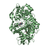
| ||||||||
| 3 | 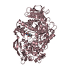
| ||||||||
| 4 | 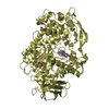
| ||||||||
| Unit cell |
|
-Components
| #1: Protein | Mass: 89198.047 Da / Num. of mol.: 4 Source method: isolated from a genetically manipulated source Source: (gene. exp.) Cryptococcus neoformans var. grubii serotype A (strain H99 / ATCC 208821 / CBS 10515 / FGSC 9487) (fungus)Strain: H99 / ATCC 208821 / CBS 10515 / FGSC 9487 / Gene: CNAG_05607 / Plasmid: ppSUMO / Production host:  #2: Chemical | ChemComp-YAJ / ~{   Mass: 319.356 Da / Num. of mol.: 4 / Source method: obtained synthetically / Formula: C16H21N3O4 / Feature type: SUBJECT OF INVESTIGATION Mass: 319.356 Da / Num. of mol.: 4 / Source method: obtained synthetically / Formula: C16H21N3O4 / Feature type: SUBJECT OF INVESTIGATION#3: Chemical | ChemComp-GOL /   Mass: 92.094 Da / Num. of mol.: 5 / Source method: obtained synthetically / Formula: C3H8O3 Mass: 92.094 Da / Num. of mol.: 5 / Source method: obtained synthetically / Formula: C3H8O3#4: Chemical | ChemComp-MG / |   Mass: 24.305 Da / Num. of mol.: 1 / Source method: obtained synthetically / Formula: Mg Mass: 24.305 Da / Num. of mol.: 1 / Source method: obtained synthetically / Formula: Mg#5: Water | ChemComp-HOH / |  Mass: 18.015 Da / Num. of mol.: 1775 / Source method: isolated from a natural source / Formula: H2O Mass: 18.015 Da / Num. of mol.: 1775 / Source method: isolated from a natural source / Formula: H2OHas ligand of interest | Y | |
|---|
-Experimental details
-Experiment
| Experiment | Method: X-RAY DIFFRACTION / Number of used crystals: 1 |
|---|
- Sample preparation
| Crystal | Density Matthews: 2.39 Å3/Da / Density % sol: 48.5 % |
|---|---|
| Crystal grow | Temperature: 293 K / Method: vapor diffusion, hanging drop / pH: 7.5 / Details: PEG8000, MgCl2, Tris 7.5, hit 9 |
-Data collection
| Diffraction | Mean temperature: 150 K / Serial crystal experiment: N | ||||||||||||||||||||||||||||||
|---|---|---|---|---|---|---|---|---|---|---|---|---|---|---|---|---|---|---|---|---|---|---|---|---|---|---|---|---|---|---|---|
| Diffraction source | Source: SYNCHROTRON / Site: APS / Beamline: 23-ID-B / Wavelength: 1.0331 Å | ||||||||||||||||||||||||||||||
| Detector | Type: DECTRIS EIGER2 X 16M / Detector: PIXEL / Date: Oct 23, 2019 | ||||||||||||||||||||||||||||||
| Radiation | Protocol: SINGLE WAVELENGTH / Monochromatic (M) / Laue (L): M / Scattering type: x-ray | ||||||||||||||||||||||||||||||
| Radiation wavelength | Wavelength: 1.0331 Å / Relative weight: 1 | ||||||||||||||||||||||||||||||
| Reflection | Resolution: 2.32→43.3 Å / Num. obs: 144484 / % possible obs: 99.8 % / Redundancy: 3.3 % / CC1/2: 0.989 / Rmerge(I) obs: 0.152 / Rpim(I) all: 0.098 / Rrim(I) all: 0.181 / Χ2: 0.82 / Net I/σ(I): 6.5 | ||||||||||||||||||||||||||||||
| Reflection shell | Diffraction-ID: 1
|
- Processing
| Software |
| |||||||||||||||||||||||||||||||||||||||||||||||||||||||||||||||||||||||||||||||||||||||||||||||||||||||||||||||||||||||||||||||||||||||||||||||||||||||||||||||||||||||||||||||||||||||||||||||||||||||||||||||||||||||||
|---|---|---|---|---|---|---|---|---|---|---|---|---|---|---|---|---|---|---|---|---|---|---|---|---|---|---|---|---|---|---|---|---|---|---|---|---|---|---|---|---|---|---|---|---|---|---|---|---|---|---|---|---|---|---|---|---|---|---|---|---|---|---|---|---|---|---|---|---|---|---|---|---|---|---|---|---|---|---|---|---|---|---|---|---|---|---|---|---|---|---|---|---|---|---|---|---|---|---|---|---|---|---|---|---|---|---|---|---|---|---|---|---|---|---|---|---|---|---|---|---|---|---|---|---|---|---|---|---|---|---|---|---|---|---|---|---|---|---|---|---|---|---|---|---|---|---|---|---|---|---|---|---|---|---|---|---|---|---|---|---|---|---|---|---|---|---|---|---|---|---|---|---|---|---|---|---|---|---|---|---|---|---|---|---|---|---|---|---|---|---|---|---|---|---|---|---|---|---|---|---|---|---|---|---|---|---|---|---|---|---|---|---|---|---|---|---|---|---|
| Refinement | Method to determine structure: MOLECULAR REPLACEMENT Starting model: 7LPO Resolution: 2.32→43.3 Å / SU ML: 0.27 / Cross valid method: THROUGHOUT / σ(F): 0 / Phase error: 24.54 / Stereochemistry target values: ML
| |||||||||||||||||||||||||||||||||||||||||||||||||||||||||||||||||||||||||||||||||||||||||||||||||||||||||||||||||||||||||||||||||||||||||||||||||||||||||||||||||||||||||||||||||||||||||||||||||||||||||||||||||||||||||
| Solvent computation | Shrinkage radii: 0.9 Å / VDW probe radii: 1.11 Å / Solvent model: FLAT BULK SOLVENT MODEL | |||||||||||||||||||||||||||||||||||||||||||||||||||||||||||||||||||||||||||||||||||||||||||||||||||||||||||||||||||||||||||||||||||||||||||||||||||||||||||||||||||||||||||||||||||||||||||||||||||||||||||||||||||||||||
| Displacement parameters | Biso max: 141.81 Å2 / Biso mean: 38.3453 Å2 / Biso min: 17.22 Å2 | |||||||||||||||||||||||||||||||||||||||||||||||||||||||||||||||||||||||||||||||||||||||||||||||||||||||||||||||||||||||||||||||||||||||||||||||||||||||||||||||||||||||||||||||||||||||||||||||||||||||||||||||||||||||||
| Refinement step | Cycle: final / Resolution: 2.32→43.3 Å
| |||||||||||||||||||||||||||||||||||||||||||||||||||||||||||||||||||||||||||||||||||||||||||||||||||||||||||||||||||||||||||||||||||||||||||||||||||||||||||||||||||||||||||||||||||||||||||||||||||||||||||||||||||||||||
| LS refinement shell | Refine-ID: X-RAY DIFFRACTION / Rfactor Rfree error: 0 / Total num. of bins used: 30
| |||||||||||||||||||||||||||||||||||||||||||||||||||||||||||||||||||||||||||||||||||||||||||||||||||||||||||||||||||||||||||||||||||||||||||||||||||||||||||||||||||||||||||||||||||||||||||||||||||||||||||||||||||||||||
| Refinement TLS params. | Method: refined / Origin x: -0.9483 Å / Origin y: 9.7018 Å / Origin z: 30.7549 Å
| |||||||||||||||||||||||||||||||||||||||||||||||||||||||||||||||||||||||||||||||||||||||||||||||||||||||||||||||||||||||||||||||||||||||||||||||||||||||||||||||||||||||||||||||||||||||||||||||||||||||||||||||||||||||||
| Refinement TLS group |
|
