 Movie
Movie Controller

[English] 日本語
 Yorodumi
- PDB-7ljv: X-ray radiation damage series on Thaumatin at 277K, crystal struc... -
+ Open data

- Basic information
| Entry | Database: PDB / ID: 7ljv | ||||||
|---|---|---|---|---|---|---|---|
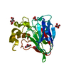
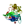
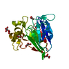
| Title | X-ray radiation damage series on Thaumatin at 277K, crystal structure, dataset 4 | ||||||
 Components Components | Thaumatin I | ||||||
 Keywords Keywords | PLANT PROTEIN / radiation damage / conformational heterogeneity | ||||||
| Function / homology |  Function and homology information Function and homology information | ||||||
| Biological species |  Thaumatococcus daniellii (katemfe) Thaumatococcus daniellii (katemfe) | ||||||
| Method |  X-RAY DIFFRACTION / SYNCHROTRON / MOLECULAR REPLACEMENT / Resolution: 1.48 Å X-RAY DIFFRACTION / SYNCHROTRON / MOLECULAR REPLACEMENT / Resolution: 1.48 Å | ||||||
 Authors Authors | Yabukarski, F. / Doukov, T. / Herschlag, D. | ||||||
| Funding support |  United States, 1items United States, 1items
| ||||||
 Citation Citation | Journal: Acta Crystallogr D Struct Biol / Year: 2022 Title: Evaluating the impact of X-ray damage on conformational heterogeneity in room-temperature (277 K) and cryo-cooled protein crystals. Authors: Yabukarski, F. / Doukov, T. / Mokhtari, D.A. / Du, S. / Herschlag, D. | ||||||
| History |
|
- Structure visualization
| Structure viewer | Molecule: MolmilJmol/JSmol |
|---|
- Downloads & links
-Download
| PDBx/mmCIF format | 7ljv.cif.gz | 165.2 KB | Display | PDBx/mmCIF format |
|---|---|---|---|---|
| PDB format | pdb7ljv.ent.gz | 111.3 KB | Display | PDB format |
| PDBx/mmJSON format | 7ljv.json.gz | Tree view | PDBx/mmJSON format | |
| Others |  Other downloads Other downloads |
-Validation report
| Arichive directory | https://data.pdbj.org/pub/pdb/validation_reports/lj/7ljvftp://data.pdbj.org/pub/pdb/validation_reports/lj/7ljv | HTTPS FTP |
|---|
-Related structure data
| Related structure data | 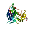 7lfgC  7ljwC  7ljzC 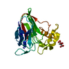 7lk5C 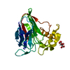 7lk6C  7llpC 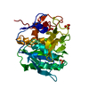 7ln7C 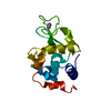 7ln8C 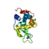 7ln9C  7lnbC 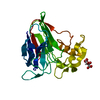 7lncC  7lndC  7loqC 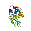 7lorC  7lp6C  7lplC  7lpmC 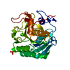 7lptC 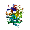 7lpuC 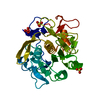 7lpvC 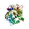 7lq8C  7lq9C  7lqaC  7lqbC  7lqcC  7ltdC  7ltiC  7ltvC  7lu0C  7lu1C  7lu2C  7lu3C  1rqwS C: citing same article ( S: Starting model for refinement |
|---|---|
| Similar structure data |


-Links
 PDBj
PDBj

- Assembly
| Deposited unit | 
| ||||||||||||
|---|---|---|---|---|---|---|---|---|---|---|---|---|---|
| 1 |
| ||||||||||||
| Unit cell |
|
-Components
| #1: Protein | Mass: 22243.119 Da / Num. of mol.: 1 / Source method: isolated from a natural source / Source: (natural) Thaumatococcus daniellii (katemfe) / References: UniProt: P02883 |
|---|---|
| #2: Chemical | ChemComp-TLA /   Mass: 150.087 Da / Num. of mol.: 1 / Source method: obtained synthetically / Formula: C4H6O6 Mass: 150.087 Da / Num. of mol.: 1 / Source method: obtained synthetically / Formula: C4H6O6 |
| #3: Water | ChemComp-HOH /  Mass: 18.015 Da / Num. of mol.: 143 / Source method: isolated from a natural source / Formula: H2O Mass: 18.015 Da / Num. of mol.: 143 / Source method: isolated from a natural source / Formula: H2O |
| Has ligand of interest | N |
| Has protein modification | Y |
| Sequence details | Thaumatin was purchased from Sigma and is a mixture of thaumatin I and thaumatin II and both are ...Thaumatin was purchased from Sigma and is a mixture of thaumatin I and thaumatin II and both are most likely present in the crystal. While thaumatin I (P02883) has an N at position 68 (precursor numbering), thaumatin II (P02884) has a K at the same position. Modeling either N or K is justified, in this case K explains the electron density better (better fit to map and better statistics). |
-Experimental details
-Experiment
| Experiment | Method: X-RAY DIFFRACTION / Number of used crystals: 1 |
|---|
- Sample preparation
| Crystal | Density Matthews: 2.94 Å3/Da / Density % sol: 58.19 % |
|---|---|
| Crystal grow | Temperature: 293 K / Method: vapor diffusion, sitting drop / pH: 6.6 Details: 24% Na K Tartrate (w/v), 15% ethylene glycol (v/v), and 0.1 M BisTris Propane |
-Data collection
| Diffraction | Mean temperature: 277 K / Serial crystal experiment: N |
|---|---|
| Diffraction source | Source: SYNCHROTRON / Site: SSRL / Beamline: BL9-2 / Wavelength: 0.88557 Å |
| Detector | Type: DECTRIS PILATUS 6M / Detector: PIXEL / Date: Nov 28, 2018 |
| Radiation | Protocol: SINGLE WAVELENGTH / Monochromatic (M) / Laue (L): M / Scattering type: x-ray |
| Radiation wavelength | Wavelength: 0.88557 Å / Relative weight: 1 |
| Reflection | Resolution: 1.48→38.29 Å / Num. obs: 45300 / % possible obs: 99.9 % / Redundancy: 8.6 % / Biso Wilson estimate: 19.21 Å2 / Rmerge(I) obs: 0.186 / Rpim(I) all: 0.066 / Net I/σ(I): 9.1 |
| Reflection shell | Resolution: 1.48→1.51 Å / Redundancy: 8 % / Rmerge(I) obs: 9.56 / Mean I/σ(I) obs: 0.6 / Num. unique obs: 2155 / Rpim(I) all: 3.509 / % possible all: 97.9 |
- Processing
| Software |
| |||||||||||||||||||||||||||||||||||||||||||||||||||||||||||||||||||||||||||||||||||||||||||||||||||||||||||||||||||||||
|---|---|---|---|---|---|---|---|---|---|---|---|---|---|---|---|---|---|---|---|---|---|---|---|---|---|---|---|---|---|---|---|---|---|---|---|---|---|---|---|---|---|---|---|---|---|---|---|---|---|---|---|---|---|---|---|---|---|---|---|---|---|---|---|---|---|---|---|---|---|---|---|---|---|---|---|---|---|---|---|---|---|---|---|---|---|---|---|---|---|---|---|---|---|---|---|---|---|---|---|---|---|---|---|---|---|---|---|---|---|---|---|---|---|---|---|---|---|---|---|---|
| Refinement | Method to determine structure: MOLECULAR REPLACEMENT Starting model: 1RQW Resolution: 1.48→37.82 Å / SU ML: 0.1625 / Cross valid method: FREE R-VALUE / σ(F): 1.34 / Phase error: 17.3809 Stereochemistry target values: GeoStd + Monomer Library + CDL v1.2
| |||||||||||||||||||||||||||||||||||||||||||||||||||||||||||||||||||||||||||||||||||||||||||||||||||||||||||||||||||||||
| Solvent computation | Shrinkage radii: 0.9 Å / VDW probe radii: 1.11 Å / Solvent model: FLAT BULK SOLVENT MODEL | |||||||||||||||||||||||||||||||||||||||||||||||||||||||||||||||||||||||||||||||||||||||||||||||||||||||||||||||||||||||
| Displacement parameters | Biso mean: 26.59 Å2 | |||||||||||||||||||||||||||||||||||||||||||||||||||||||||||||||||||||||||||||||||||||||||||||||||||||||||||||||||||||||
| Refinement step | Cycle: LAST / Resolution: 1.48→37.82 Å
| |||||||||||||||||||||||||||||||||||||||||||||||||||||||||||||||||||||||||||||||||||||||||||||||||||||||||||||||||||||||
| Refine LS restraints |
| |||||||||||||||||||||||||||||||||||||||||||||||||||||||||||||||||||||||||||||||||||||||||||||||||||||||||||||||||||||||
| LS refinement shell |
|
