 Movie
Movie Controller


+ Open data

- Basic information
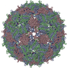
| Entry | Database: PDB / ID: 7lhd | |||||||||||||||
|---|---|---|---|---|---|---|---|---|---|---|---|---|---|---|---|---|
| Title | The complete model of phage Qbeta virion | |||||||||||||||
 Components Components |
| |||||||||||||||
 Keywords Keywords | VIRUS/RNA / Bacteriophage / Virus / Virion / VIRUS-RNA complex | |||||||||||||||
| Function / homology |  Function and homology information Function and homology informationsymbiont-mediated suppression of host cell wall biogenesis / symbiont-mediated suppression of host peptidoglycan biosynthetic process / viral release via suppression of host peptidoglycan biosynthetic process / virion attachment to host cell pilus / T=3 icosahedral viral capsid / adhesion receptor-mediated virion attachment to host cell / viral release from host cell by cytolysis / translation repressor activity / virion component / structural molecule activity / RNA binding Similarity search - Function | |||||||||||||||
| Biological species |  Escherichia virus QbetaEscherichia phage Qbeta (virus) Escherichia virus QbetaEscherichia phage Qbeta (virus) | |||||||||||||||
| Method | ELECTRON MICROSCOPY / single particle reconstruction / cryo EM / Resolution: 4.6 Å | |||||||||||||||
 Authors Authors | Chang, J.Y. / Zhang, J. | |||||||||||||||
| Funding support |  United States, 4items United States, 4items
| |||||||||||||||
 Citation Citation | Journal: Viruses / Year: 2022 Title: Structural Assembly of Qβ Virion and Its Diverse Forms of Virus-like Particles. Authors: Jeng-Yih Chang / Karl V Gorzelnik / Jirapat Thongchol / Junjie Zhang / Abstract: The coat proteins (CPs) of single-stranded RNA bacteriophages (ssRNA phages) directly assemble around the genomic RNA (gRNA) to form a near-icosahedral capsid with a single maturation protein (Mat) ...The coat proteins (CPs) of single-stranded RNA bacteriophages (ssRNA phages) directly assemble around the genomic RNA (gRNA) to form a near-icosahedral capsid with a single maturation protein (Mat) that binds the gRNA and interacts with the retractile pilus during infection of the host. Understanding the assembly of ssRNA phages is essential for their use in biotechnology, such as RNA protection and delivery. Here, we present the complete gRNA model of the ssRNA phage Qβ, revealing that the 3' untranslated region binds to the Mat and the 4127 nucleotides fold domain-by-domain, and is connected through long-range RNA-RNA interactions, such as kissing loops. Thirty-three operator-like RNA stem-loops are located and primarily interact with the asymmetric A/B CP-dimers, suggesting a pathway for the assembly of the virions. Additionally, we have discovered various forms of the virus-like particles (VLPs), including the canonical = 3 icosahedral, larger = 4 icosahedral, prolate, oblate forms, and a small prolate form elongated along the 3-fold axis. These particles are all produced during a normal infection, as well as when overexpressing the CPs. When overexpressing the shorter RNA fragments encoding only the CPs, we observed an increased percentage of the smaller VLPs, which may be sufficient to encapsidate a shorter RNA. | |||||||||||||||
| History |
|
- Structure visualization
| Movie |
Movie viewer |
|---|---|
| Structure viewer | Molecule: MolmilJmol/JSmol |
 UCSF Chimera
- Downloads & links
-Download
| PDBx/mmCIF format | 7lhd.cif.gz | 5.7 MB | Display | PDBx/mmCIF format |
|---|---|---|---|---|
| PDB format | pdb7lhd.ent.gz | Display | PDB format | |
| PDBx/mmJSON format | 7lhd.json.gz | Tree view | PDBx/mmJSON format | |
| Others |  Other downloads Other downloads |
-Validation report
| Arichive directory | https://data.pdbj.org/pub/pdb/validation_reports/lh/7lhdftp://data.pdbj.org/pub/pdb/validation_reports/lh/7lhd | HTTPS FTP |
|---|
-Related structure data
| Related structure data |  23336MC  7lgeC  7lgfC  7lggC  7lghC M: map data used to model this data C: citing same article ( |
|---|---|
| Similar structure data |


-Links
 PDBj
PDBj


- Assembly
| Deposited unit | 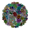
|
|---|---|
| 1 |
|
-Components
| #1: RNA chain | Mass: 1352016.625 Da / Num. of mol.: 1 / Source method: isolated from a natural source / Source: (natural) Escherichia virus Qbeta / References: GenBank: 668235487 | ||
|---|---|---|---|
| #2: Protein | Mass: 48613.078 Da / Num. of mol.: 1 / Source method: isolated from a natural source / Source: (natural) Escherichia phage Qbeta (virus) / References: UniProt: Q8LTE2 | ||
| #3: Protein | Mass: 14268.071 Da / Num. of mol.: 180 / Source method: isolated from a natural source / Source: (natural) Escherichia phage Qbeta (virus) / References: UniProt: P03615Has protein modification | Y | |
-Experimental details
-Experiment
| Experiment | Method: ELECTRON MICROSCOPY |
|---|---|
| EM experiment | Aggregation state: PARTICLE / 3D reconstruction method: single particle reconstruction |
- Sample preparation
| Component | Name: Escherichia virus Qbeta / Type: VIRUS / Entity ID: all / Source: NATURAL |
|---|---|
| Source (natural) | Organism: Escherichia virus Qbeta |
| Details of virus | Empty: NO / Enveloped: NO / Isolate: SPECIES / Type: VIRION |
| Buffer solution | pH: 7 |
| Specimen | Embedding applied: NO / Shadowing applied: NO / Staining applied: NO / Vitrification applied: YES |
| Vitrification | Cryogen name: ETHANE |
- Electron microscopy imaging
| Experimental equipment |  Model: Tecnai F20 / Image courtesy: FEI Company | |||||||||||||||||||||
|---|---|---|---|---|---|---|---|---|---|---|---|---|---|---|---|---|---|---|---|---|---|---|
| EM imaging |
| |||||||||||||||||||||
| Image recording |
|
- Processing
| CTF correction | Type: PHASE FLIPPING AND AMPLITUDE CORRECTION |
|---|---|
| 3D reconstruction | Resolution: 4.6 Å / Resolution method: FSC 0.143 CUT-OFF / Num. of particles: 86081 / Symmetry type: POINT |
