| Entry | Database: PDB / ID: 7ld9
|
|---|
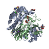
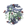
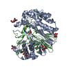
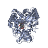
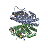
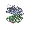
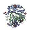
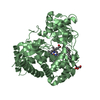
| Title | Structure of human GGT1 in complex with ABBA |
|---|
 Components Components | (Glutathione hydrolase 1 ...) x 2 |
|---|
 Keywords Keywords | HYDROLASE/HYDROLASE INHIBITOR / substrate-enzyme complex / NTN-hydrolase family / glycoprotein / N-glycosylation / cell surface / HYDROLASE-HYDROLASE INHIBITOR complex / ABBA |
|---|
| Function / homology |  Function and homology information Function and homology information
leukotriene-C4 hydrolase / peptidyltransferase activity / leukotriene-C(4) hydrolase activity / leukotriene D4 biosynthetic process / Defective GGT1 causes GLUTH / Defective GGT1 in aflatoxin detoxification causes GLUTH / Glutathione synthesis and recycling / LTC4-CYSLTR mediated IL4 production / gamma-glutamyltransferase / glutathione gamma-glutamate hydrolase ...leukotriene-C4 hydrolase / peptidyltransferase activity / leukotriene-C(4) hydrolase activity / leukotriene D4 biosynthetic process / Defective GGT1 causes GLUTH / Defective GGT1 in aflatoxin detoxification causes GLUTH / Glutathione synthesis and recycling / LTC4-CYSLTR mediated IL4 production / gamma-glutamyltransferase / glutathione gamma-glutamate hydrolase / peptide modification / glutathione gamma-glutamate hydrolase / leukotriene C4 gamma-glutamyl transferase activity / glutathione catabolic process / glutamate metabolic process / glutathione biosynthetic process / L-cysteine biosynthetic process / leukotriene metabolic process / Aflatoxin activation and detoxification / Synthesis of Leukotrienes (LT) and Eoxins (EX) / Paracetamol ADME / amino acid metabolic process / regulation of immune system process / zymogen activation / fatty acid metabolic process / regulation of inflammatory response / spermatogenesis / proteolysis / extracellular space / extracellular exosome / plasma membraneSimilarity search - Function : / Gamma-glutamyltranspeptidase / Gamma-glutamyltranspeptidase signature. / Gamma-glutamyltranspeptidase / Gamma-glutamyltranspeptidase, large subunit, C-terminal domain / Gamma-glutamyltranspeptidase, small subunit / Nucleophile aminohydrolases, N-terminalSimilarity search - Domain/homology |
|---|
| Biological species |  Homo sapiens (human) Homo sapiens (human) |
|---|
| Method |  X-RAY DIFFRACTION / SYNCHROTRON / FOURIER SYNTHESIS / Resolution: 1.42 Å X-RAY DIFFRACTION / SYNCHROTRON / FOURIER SYNTHESIS / Resolution: 1.42 Å |
|---|
 Authors Authors | Terzyan, S.S. / Hanigan, M. |
|---|
| Funding support |  United States, 1items United States, 1items | Organization | Grant number | Country |
|---|
| National Institutes of Health/National Institute of General Medical Sciences (NIH/NIGMS) | R01GM125952 | United States |
|
|---|
 Citation Citation | Journal: Bioorg.Med.Chem. / Year: 2022
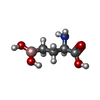
Title: Design and evaluation of novel analogs of 2-amino-4-boronobutanoic acid (ABBA) as inhibitors of human gamma-glutamyl transpeptidase.
Authors: Nguyen, L. / Schultz, D.C. / Terzyan, S.S. / Rezaei, M. / Songb, J. / Li, C. / You, Y. / Hanigan, M.H. |
|---|
| History | | Deposition | Jan 12, 2021 | Deposition site: RCSB / Processing site: RCSB |
|---|
| Revision 1.0 | Feb 23, 2022 | Provider: repository / Type: Initial release |
|---|
| Revision 1.1 | Mar 8, 2023 | Group: Database references / Category: citation / citation_author
Item: _citation.country / _citation.journal_abbrev ..._citation.country / _citation.journal_abbrev / _citation.journal_id_ASTM / _citation.journal_id_CSD / _citation.journal_id_ISSN / _citation.journal_volume / _citation.page_first / _citation.page_last / _citation.pdbx_database_id_DOI / _citation.pdbx_database_id_PubMed / _citation.title / _citation.year |
|---|
| Revision 1.2 | Oct 25, 2023 | Group: Data collection / Refinement description
Category: chem_comp_atom / chem_comp_bond / pdbx_initial_refinement_model |
|---|
| Revision 1.3 | Nov 6, 2024 | Group: Structure summary / Category: pdbx_entry_details / pdbx_modification_feature / Item: _pdbx_entry_details.has_protein_modification |
|---|
|
|---|
 Movie
Movie Controller


 Open data
 Basic information
 Structure visualization
 Downloads & links
 Other downloads


 PDBj
PDBj


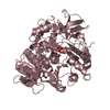
 Assembly
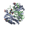
 Komagataella pastoris (fungus) / Strain (production host): X-33
 Type: D-saccharide, beta linking / Mass: 221.208 Da / Num. of mol.: 6 / Source method: obtained synthetically / Formula: C8H15NO6


 Mass: 35.453 Da / Num. of mol.: 2 / Source method: obtained synthetically / Formula: Cl
 Mass: 22.990 Da / Num. of mol.: 1 / Source method: obtained synthetically / Formula: Na
 Mass: 238.305 Da / Num. of mol.: 3 / Source method: isolated from a natural source / Formula: C8H18N2O4S / Comment: pH buffer*YM
 Mass: 146.937 Da / Num. of mol.: 1 / Source method: obtained synthetically / Formula: C4H10BNO4
 Mass: 146.937 Da / Num. of mol.: 1 / Source method: obtained synthetically / Formula: C4H10BNO4
 Sample preparation
 Processing