 Movie
Movie Controller


+ Open data

- Basic information
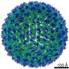
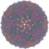
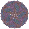
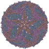
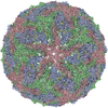
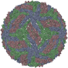
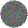
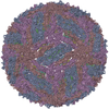
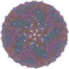
| Entry | Database: PDB / ID: 7lch | |||||||||
|---|---|---|---|---|---|---|---|---|---|---|
| Title | The mature Usutu SAAR-1776, Model B | |||||||||
 Components Components |
| |||||||||
 Keywords Keywords | VIRUS / Flavivirus / envelope glycoprotein | |||||||||
| Function / homology |  Function and homology information Function and homology informationsymbiont-mediated suppression of host JAK-STAT cascade via inhibition of STAT2 activity / viral capsid / ribonucleoside triphosphate phosphatase activity / double-stranded RNA binding / methyltransferase cap1 activity / mRNA 5'-cap (guanine-N7-)-methyltransferase activity / RNA helicase activity / protein dimerization activity / symbiont-mediated suppression of host innate immune response / host cell perinuclear region of cytoplasm ...symbiont-mediated suppression of host JAK-STAT cascade via inhibition of STAT2 activity / viral capsid / ribonucleoside triphosphate phosphatase activity / double-stranded RNA binding / methyltransferase cap1 activity / mRNA 5'-cap (guanine-N7-)-methyltransferase activity / RNA helicase activity / protein dimerization activity / symbiont-mediated suppression of host innate immune response / host cell perinuclear region of cytoplasm / host cell endoplasmic reticulum membrane / symbiont-mediated suppression of host type I interferon-mediated signaling pathway / serine-type endopeptidase activity / viral RNA genome replication / RNA-directed RNA polymerase activity / fusion of virus membrane with host endosome membrane / symbiont entry into host cell / virion attachment to host cell / host cell nucleus / virion membrane / structural molecule activity / proteolysis / extracellular region / ATP binding / metal ion binding Similarity search - Function | |||||||||
| Biological species |  Usutu virus Usutu virus | |||||||||
| Method | ELECTRON MICROSCOPY / single particle reconstruction / cryo EM / Resolution: 2.35 Å | |||||||||
 Authors Authors | Khare, B. / Klose, T. / Fang, Q. / Kuhn, R. | |||||||||
| Funding support |  United States, 2items United States, 2items
| |||||||||
 Citation Citation | Journal: Proc Natl Acad Sci U S A / Year: 2021 Title: Structure of Usutu virus SAAR-1776 displays fusion loop asymmetry. Authors: Baldeep Khare / Thomas Klose / Qianglin Fang / Michael G Rossmann / Richard J Kuhn / Abstract: Usutu virus (USUV) is an emerging arbovirus in Europe that has been increasingly identified in asymptomatic humans and donated blood samples and is a cause of increased incidents of neuroinvasive ...Usutu virus (USUV) is an emerging arbovirus in Europe that has been increasingly identified in asymptomatic humans and donated blood samples and is a cause of increased incidents of neuroinvasive human disease. Treatment or prevention options for USUV disease are currently nonexistent, the result of a lack of understanding of the fundamental elements of USUV pathogenesis. Here, we report two structures of the mature USUV virus, determined at a resolution of 2.4 Å, using single-particle cryogenic electron microscopy. Mature USUV is an icosahedral shell of 180 copies of envelope (E) and membrane (M) proteins arranged in the classic herringbone pattern. However, unlike previous reports of flavivirus structures, we observe virus subpopulations and differences in the fusion loop disulfide bond. Presence of a second, unique E glycosylation site could elucidate host interactions, contributing to the broad USUV tissue tropism. The structures provide a basis for exploring USUV interactions with glycosaminoglycans and lectins, the role of the RGD motif as a receptor, and the inability of West Nile virus therapeutic antibody E16 to neutralize the mature USUV strain SAAR-1776. Finally, we identify three lipid binding sites and predict key residues that likely participate in virus stability and flexibility during membrane fusion. Our findings provide a framework for the development of USUV therapeutics and expand the current knowledge base of flavivirus biology. | |||||||||
| History |
|
- Structure visualization
| Movie |
Movie viewer |
|---|---|
| Structure viewer | Molecule: MolmilJmol/JSmol |
- Downloads & links
-Download
| PDBx/mmCIF format | 7lch.cif.gz | 310.2 KB | Display | PDBx/mmCIF format |
|---|---|---|---|---|
| PDB format | pdb7lch.ent.gz | 255.8 KB | Display | PDB format |
| PDBx/mmJSON format | 7lch.json.gz | Tree view | PDBx/mmJSON format | |
| Others |  Other downloads Other downloads |
-Validation report
| Arichive directory | https://data.pdbj.org/pub/pdb/validation_reports/lc/7lchftp://data.pdbj.org/pub/pdb/validation_reports/lc/7lch | HTTPS FTP |
|---|
-Related structure data
| Related structure data | 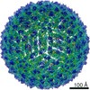 23273MC 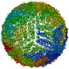 7lcgC M: map data used to model this data C: citing same article ( |
|---|---|
| Similar structure data |

-Links
 PDBj
PDBj

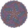
- Assembly
| Deposited unit | 
|
|---|---|
| 1 | x 60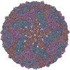
|
| 2 |
|
| 3 | x 5
|
| 4 | x 6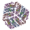
|
| 5 | 
|
| Symmetry | Point symmetry: (Schoenflies symbol: I (icosahedral)) |
-Components
| #1: Protein | Mass: 53623.812 Da / Num. of mol.: 3 / Fragment: UNP residues 294-793 Source method: isolated from a genetically manipulated source Source: (gene. exp.) Usutu virus / Strain: SAAR-1776 / Cell line (production host): Vero / Production host:  Chlorocebus aethiops (grivet monkey) / References: UniProt: Q5WPU4 Chlorocebus aethiops (grivet monkey) / References: UniProt: Q5WPU4#2: Protein | Mass: 8333.710 Da / Num. of mol.: 3 / Fragment: UNP residues 219-293 Source method: isolated from a genetically manipulated source Source: (gene. exp.) Usutu virus / Strain: SAAR-1776 / Cell line (production host): Vero / Production host: Chlorocebus aethiops (grivet monkey) / References: UniProt: A0A0H3U5P6#3: Chemical | 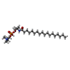  Mass: 537.733 Da / Num. of mol.: 3 / Source method: obtained synthetically / Formula: C27H58N2O6P / Feature type: SUBJECT OF INVESTIGATION Mass: 537.733 Da / Num. of mol.: 3 / Source method: obtained synthetically / Formula: C27H58N2O6P / Feature type: SUBJECT OF INVESTIGATION#4: Chemical |   Mass: 763.100 Da / Num. of mol.: 3 / Source method: obtained synthetically / Formula: C42H85NO8P / Feature type: SUBJECT OF INVESTIGATION / Comment: phospholipid*YM Mass: 763.100 Da / Num. of mol.: 3 / Source method: obtained synthetically / Formula: C42H85NO8P / Feature type: SUBJECT OF INVESTIGATION / Comment: phospholipid*YM#5: Sugar | ChemComp-NAG /   Type: D-saccharide, beta linking / Mass: 221.208 Da / Num. of mol.: 6 / Source method: obtained synthetically / Formula: C8H15NO6 / Feature type: SUBJECT OF INVESTIGATION Type: D-saccharide, beta linking / Mass: 221.208 Da / Num. of mol.: 6 / Source method: obtained synthetically / Formula: C8H15NO6 / Feature type: SUBJECT OF INVESTIGATIONHas ligand of interest | Y | Has protein modification | Y | |
|---|
-Experimental details
-Experiment
| Experiment | Method: ELECTRON MICROSCOPY |
|---|---|
| EM experiment | Aggregation state: PARTICLE / 3D reconstruction method: single particle reconstruction |
- Sample preparation
| Component | Name: Usutu virus / Type: VIRUS / Details: Virus purified using Vero cells. / Entity ID: #1-#2 / Source: RECOMBINANT | ||||||||||||||||||||
|---|---|---|---|---|---|---|---|---|---|---|---|---|---|---|---|---|---|---|---|---|---|
| Source (natural) | Organism: Usutu virus / Strain: SAAR-1776 | ||||||||||||||||||||
| Source (recombinant) | Organism: Chlorocebus aethiops (grivet monkey) / Cell: Vero cells | ||||||||||||||||||||
| Details of virus | Empty: NO / Enveloped: YES / Isolate: STRAIN / Type: VIRION | ||||||||||||||||||||
| Natural host | Organism: Turdus merula | ||||||||||||||||||||
| Buffer solution | pH: 8 / Details: 20 mM Tris, 120 mM NaCl, 1 mM EDTA, pH 8.0 | ||||||||||||||||||||
| Buffer component |
| ||||||||||||||||||||
| Specimen | Embedding applied: NO / Shadowing applied: NO / Staining applied: NO / Vitrification applied: YES | ||||||||||||||||||||
| Specimen support | Grid material: COPPER / Grid type: PELCO Ultrathin Carbon with Lacey Carbon | ||||||||||||||||||||
| Vitrification | Instrument: GATAN CRYOPLUNGE 3 / Cryogen name: ETHANE |
- Electron microscopy imaging
| Experimental equipment |  Model: Titan Krios / Image courtesy: FEI Company |
|---|---|
| Microscopy | Model: FEI TITAN KRIOS |
| Electron gun | Electron source:  FIELD EMISSION GUN / Accelerating voltage: 300 kV / Illumination mode: FLOOD BEAM FIELD EMISSION GUN / Accelerating voltage: 300 kV / Illumination mode: FLOOD BEAM |
| Electron lens | Mode: BRIGHT FIELD / Nominal magnification: 64000 X / Nominal defocus max: 2000 nm / Nominal defocus min: 800 nm / Cs: 2.7 mm / C2 aperture diameter: 100 µm / Alignment procedure: COMA FREE |
| Specimen holder | Cryogen: NITROGEN / Specimen holder model: FEI TITAN KRIOS AUTOGRID HOLDER |
| Image recording | Average exposure time: 2.08 sec. / Electron dose: 23.68 e/Å2 / Detector mode: SUPER-RESOLUTION / Film or detector model: GATAN K3 BIOQUANTUM (6k x 4k) / Num. of grids imaged: 1 / Num. of real images: 3689 |
| EM imaging optics | Energyfilter name: GIF Bioquantum / Energyfilter slit width: 20 eV |
| Image scans | Movie frames/image: 40 |
- Processing
| Software | Name: PHENIX / Version: 1.19_4085: / Classification: refinement | ||||||||||||||||||||||||||||||
|---|---|---|---|---|---|---|---|---|---|---|---|---|---|---|---|---|---|---|---|---|---|---|---|---|---|---|---|---|---|---|---|
| EM software |
| ||||||||||||||||||||||||||||||
| CTF correction | Type: PHASE FLIPPING ONLY | ||||||||||||||||||||||||||||||
| Symmetry | Point symmetry: I (icosahedral) | ||||||||||||||||||||||||||||||
| 3D reconstruction | Resolution: 2.35 Å / Resolution method: FSC 0.143 CUT-OFF / Num. of particles: 285180 / Algorithm: FOURIER SPACE / Symmetry type: POINT | ||||||||||||||||||||||||||||||
| Atomic model building | Space: REAL | ||||||||||||||||||||||||||||||
| Refinement | Highest resolution: 2.35 Å | ||||||||||||||||||||||||||||||
| Refine LS restraints |
|
