 Movie
Movie Controller

[English] 日本語
 Yorodumi
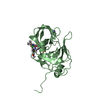
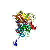
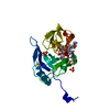
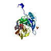
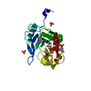
- PDB-7l7n: Crystal structure of HCV NS3/4A D168A protease in complex with NR02-59 -
+ Open data

- Basic information
| Entry | Database: PDB / ID: 7l7n | |||||||||
|---|---|---|---|---|---|---|---|---|---|---|
| Title | Crystal structure of HCV NS3/4A D168A protease in complex with NR02-59 | |||||||||
 Components Components | NS3 protease | |||||||||
 Keywords Keywords | HYDROLASE/INHIBITOR / NS3/4a Protease / Hepatitis C virus / Drug Resistance / Protease inhibitor / HYDROLASE-HYDROLASE Inhibitor complex / HYDROLASE / HYDROLASE-INHIBITOR complex | |||||||||
| Function / homology |  Function and homology information Function and homology informationsymbiont-mediated transformation of host cell / host cell membrane / serine-type peptidase activity / host cell / symbiont entry into host cell / virion attachment to host cell / virion membrane / proteolysis / metal ion binding / membrane Similarity search - Function | |||||||||
| Biological species |  Hepacivirus C Hepacivirus C | |||||||||
| Method |  X-RAY DIFFRACTION / MOLECULAR REPLACEMENT / Resolution: 1.59 Å X-RAY DIFFRACTION / MOLECULAR REPLACEMENT / Resolution: 1.59 Å | |||||||||
 Authors Authors | Zephyr, J. / Schiffer, C.A. | |||||||||
| Funding support |  United States, 2items United States, 2items
| |||||||||
 Citation Citation | Journal: J.Med.Chem. / Year: 2021 Title: Discovery of Quinoxaline-Based P1-P3 Macrocyclic NS3/4A Protease Inhibitors with Potent Activity against Drug-Resistant Hepatitis C Virus Variants. Authors: Nageswara Rao, D. / Zephyr, J. / Henes, M. / Chan, E.T. / Matthew, A.N. / Hedger, A.K. / Conway, H.L. / Saeed, M. / Newton, A. / Petropoulos, C.J. / Huang, W. / Kurt Yilmaz, N. / Schiffer, C.A. / Ali, A. | |||||||||
| History |
|
- Structure visualization
| Structure viewer | Molecule: MolmilJmol/JSmol |
|---|
- Downloads & links
-Download
| PDBx/mmCIF format | 7l7n.cif.gz | 111.7 KB | Display | PDBx/mmCIF format |
|---|---|---|---|---|
| PDB format | pdb7l7n.ent.gz | 68.5 KB | Display | PDB format |
| PDBx/mmJSON format | 7l7n.json.gz | Tree view | PDBx/mmJSON format | |
| Others |  Other downloads Other downloads |
-Validation report
| Summary document | 7l7n_validation.pdf.gz | 797.1 KB | Display | wwPDB validaton report |
|---|---|---|---|---|
| Full document | 7l7n_full_validation.pdf.gz | 801.8 KB | Display | |
| Data in XML | 7l7n_validation.xml.gz | 12.9 KB | Display | |
| Data in CIF | 7l7n_validation.cif.gz | 18.2 KB | Display | |
| Arichive directory | https://data.pdbj.org/pub/pdb/validation_reports/l7/7l7nftp://data.pdbj.org/pub/pdb/validation_reports/l7/7l7n | HTTPS FTP |
-Related structure data
| Related structure data |  7l7lC  7l7oC  7l7pC  5vojS S: Starting model for refinement C: citing same article ( |
|---|---|
| Similar structure data |


-Links
 PDBj
PDBj
- Assembly
| Deposited unit | 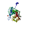
| ||||||||||||
|---|---|---|---|---|---|---|---|---|---|---|---|---|---|
| 1 |
| ||||||||||||
| Unit cell |
|
-Components
-Protein , 1 types, 1 molecules A
| #1: Protein | Mass: 23486.564 Da / Num. of mol.: 1 / Mutation: D168A Source method: isolated from a genetically manipulated source Source: (gene. exp.) Hepacivirus C / Plasmid: PET28a / Production host:  |
|---|
-Non-polymers , 5 types, 201 molecules 


| #2: Chemical | ChemComp-ZN /  Mass: 65.409 Da / Num. of mol.: 1 / Source method: obtained synthetically / Formula: Zn Mass: 65.409 Da / Num. of mol.: 1 / Source method: obtained synthetically / Formula: Zn | ||||
|---|---|---|---|---|---|
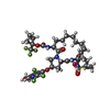
| #3: Chemical | ChemComp-XSV /  Mass: 874.846 Da / Num. of mol.: 1 / Source method: obtained synthetically / Formula: C38H44F6N6O9S / Feature type: SUBJECT OF INVESTIGATION Mass: 874.846 Da / Num. of mol.: 1 / Source method: obtained synthetically / Formula: C38H44F6N6O9S / Feature type: SUBJECT OF INVESTIGATION | ||||
| #4: Chemical |  Mass: 62.068 Da / Num. of mol.: 2 / Source method: obtained synthetically / Formula: C2H6O2 Mass: 62.068 Da / Num. of mol.: 2 / Source method: obtained synthetically / Formula: C2H6O2#5: Chemical |  Mass: 96.063 Da / Num. of mol.: 2 / Source method: obtained synthetically / Formula: SO4 Mass: 96.063 Da / Num. of mol.: 2 / Source method: obtained synthetically / Formula: SO4#6: Water | ChemComp-HOH / | Mass: 18.015 Da / Num. of mol.: 195 / Source method: isolated from a natural source / Formula: H2O |
-Details
| Has ligand of interest | Y |
|---|
-Experimental details
-Experiment
| Experiment | Method: X-RAY DIFFRACTION / Number of used crystals: 1 |
|---|
- Sample preparation
| Crystal | Density Matthews: 2.06 Å3/Da / Density % sol: 40.17 % |
|---|---|
| Crystal grow | Temperature: 298 K / Method: vapor diffusion, hanging drop / pH: 6.5 Details: 100 mM MES Buffer pH 6.5, 4% (W/V) Ammonium Sulfate, 20-26% PEG 3350 The crystals were then soaked overnight in cryogenic conditions containing inhibitor (100 mM MES Buffer pH 6.5, 4% (W/V) ...Details: 100 mM MES Buffer pH 6.5, 4% (W/V) Ammonium Sulfate, 20-26% PEG 3350 The crystals were then soaked overnight in cryogenic conditions containing inhibitor (100 mM MES Buffer pH 6.5, 4% (W/V) Ammonium Sulfate, 20-26% PEG 3350, 15% Ethylene glycol, and 10-20 mM of inhibitor in DMF) |
-Data collection
| Diffraction | Mean temperature: 100 K / Serial crystal experiment: N |
|---|---|
| Diffraction source | Source: ROTATING ANODE / Type: RIGAKU MICROMAX-007 HF / Wavelength: 1.54178 Å |
| Detector | Type: RIGAKU SATURN 944 / Detector: CCD / Date: Sep 23, 2020 |
| Radiation | Protocol: SINGLE WAVELENGTH / Monochromatic (M) / Laue (L): M / Scattering type: x-ray |
| Radiation wavelength | Wavelength: 1.54178 Å / Relative weight: 1 |
| Reflection | Resolution: 1.59→24.0555 Å / Num. obs: 26388 / % possible obs: 98.55 % / Redundancy: 4.6 % / Biso Wilson estimate: 19.22 Å2 / CC1/2: 0.996 / Net I/σ(I): 12.48 |
| Reflection shell | Resolution: 1.59→1.647 Å / Num. unique obs: 2355 / CC1/2: 0.838 |
- Processing
| Software |
| ||||||||||||||||||||||||||||||||||||||||||||||||||||||||||||||||||||||
|---|---|---|---|---|---|---|---|---|---|---|---|---|---|---|---|---|---|---|---|---|---|---|---|---|---|---|---|---|---|---|---|---|---|---|---|---|---|---|---|---|---|---|---|---|---|---|---|---|---|---|---|---|---|---|---|---|---|---|---|---|---|---|---|---|---|---|---|---|---|---|---|
| Refinement | Method to determine structure: MOLECULAR REPLACEMENT Starting model: 5VOJ Resolution: 1.59→24.05 Å / SU ML: 0.1794 / Cross valid method: FREE R-VALUE / σ(F): 1.34 / Phase error: 22.3013 Stereochemistry target values: GeoStd + Monomer Library + CDL v1.2
| ||||||||||||||||||||||||||||||||||||||||||||||||||||||||||||||||||||||
| Solvent computation | Shrinkage radii: 0.9 Å / VDW probe radii: 1.11 Å / Solvent model: FLAT BULK SOLVENT MODEL | ||||||||||||||||||||||||||||||||||||||||||||||||||||||||||||||||||||||
| Displacement parameters | Biso mean: 23.12 Å2 | ||||||||||||||||||||||||||||||||||||||||||||||||||||||||||||||||||||||
| Refinement step | Cycle: LAST / Resolution: 1.59→24.05 Å
| ||||||||||||||||||||||||||||||||||||||||||||||||||||||||||||||||||||||
| Refine LS restraints |
| ||||||||||||||||||||||||||||||||||||||||||||||||||||||||||||||||||||||
| LS refinement shell |
|
