 Movie
Movie Controller

[English] 日本語
 Yorodumi
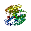
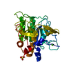
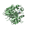
- PDB-7l1w: Unlocking the structural features for the exo-xylobiosidase activ... -
+ Open data

- Basic information
| Entry | Database: PDB / ID: 7l1w | |||||||||
|---|---|---|---|---|---|---|---|---|---|---|
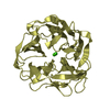
| Title | Unlocking the structural features for the exo-xylobiosidase activity of an unusual GH11 member identified in a compost-derived consortium | |||||||||
 Components Components | Exo-B-1,4-beta-xylanase | |||||||||
 Keywords Keywords | HYDROLASE / glycoside hydrolase family 11 / GH11 Exo-B-1 / 4-xylobiosidase | |||||||||
| Function / homology | Glycoside hydrolase family 11/12, catalytic domain / Jelly Rolls / Sandwich / Mainly Beta Function and homology information Function and homology information | |||||||||
| Biological species | unidentified (others) | |||||||||
| Method |  X-RAY DIFFRACTION / SYNCHROTRON / MOLECULAR REPLACEMENT / Resolution: 1.71 Å X-RAY DIFFRACTION / SYNCHROTRON / MOLECULAR REPLACEMENT / Resolution: 1.71 Å | |||||||||
 Authors Authors | Kadowaki, M.A.S. / Polikarpov, I. / Briganti, L. / Evangelista, D.E. | |||||||||
| Funding support |  Brazil, 2items Brazil, 2items
| |||||||||
 Citation Citation | Journal: Biotechnol.Bioeng. / Year: 2021 Title: Unlocking the structural features for the xylobiohydrolase activity of an unusual GH11 member identified in a compost-derived consortium. Authors: Kadowaki, M.A.S. / Briganti, L. / Evangelista, D.E. / Echevarria-Poza, A. / Tryfona, T. / Pellegrini, V.O.A. / Nakayama, D.G. / Dupree, P. / Polikarpov, I. | |||||||||
| History |
|
- Structure visualization
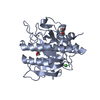
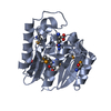
| Structure viewer | Molecule: MolmilJmol/JSmol |
|---|
- Downloads & links
-Download
| PDBx/mmCIF format | 7l1w.cif.gz | 70.1 KB | Display | PDBx/mmCIF format |
|---|---|---|---|---|
| PDB format | pdb7l1w.ent.gz | 49 KB | Display | PDB format |
| PDBx/mmJSON format | 7l1w.json.gz | Tree view | PDBx/mmJSON format | |
| Others |  Other downloads Other downloads |
-Validation report
| Summary document | 7l1w_validation.pdf.gz | 435.5 KB | Display | wwPDB validaton report |
|---|---|---|---|---|
| Full document | 7l1w_full_validation.pdf.gz | 436.4 KB | Display | |
| Data in XML | 7l1w_validation.xml.gz | 13.7 KB | Display | |
| Data in CIF | 7l1w_validation.cif.gz | 20 KB | Display | |
| Arichive directory | https://data.pdbj.org/pub/pdb/validation_reports/l1/7l1wftp://data.pdbj.org/pub/pdb/validation_reports/l1/7l1w | HTTPS FTP |
-Related structure data
| Related structure data | 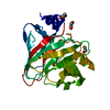 7l1yC  7l1zC 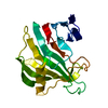 5vqjS S: Starting model for refinement C: citing same article ( |
|---|---|
| Similar structure data |


-Links
 PDBj
PDBj- Assembly
| Deposited unit | 
| ||||||||||||
|---|---|---|---|---|---|---|---|---|---|---|---|---|---|
| 1 |
| ||||||||||||
| Unit cell |
| ||||||||||||
| Components on special symmetry positions |
|
-Components
| #1: Protein | Mass: 27681.490 Da / Num. of mol.: 1 Source method: isolated from a genetically manipulated source Details: Metatranscriptome / Source: (gene. exp.) unidentified (others) / Production host:  |
|---|---|
| #2: Chemical | ChemComp-MES /   Mass: 195.237 Da / Num. of mol.: 1 / Source method: obtained synthetically / Formula: C6H13NO4S / Comment: pH buffer*YM Mass: 195.237 Da / Num. of mol.: 1 / Source method: obtained synthetically / Formula: C6H13NO4S / Comment: pH buffer*YM |
| #3: Water | ChemComp-HOH /  Mass: 18.015 Da / Num. of mol.: 242 / Source method: isolated from a natural source / Formula: H2O Mass: 18.015 Da / Num. of mol.: 242 / Source method: isolated from a natural source / Formula: H2O |
| Has ligand of interest | N |
-Experimental details
-Experiment
| Experiment | Method: X-RAY DIFFRACTION / Number of used crystals: 1 |
|---|
- Sample preparation
| Crystal | Density Matthews: 2 Å3/Da / Density % sol: 38.64 % |
|---|---|
| Crystal grow | Temperature: 291 K / Method: vapor diffusion, sitting drop / pH: 6.5 / Details: 0.1M MES pH 6.5 and 30% (w/v) PEG 4000 |
-Data collection
| Diffraction | Mean temperature: 100 K / Serial crystal experiment: N |
|---|---|
| Diffraction source | Source: SYNCHROTRON / Site: LNLS / Beamline: W01B-MX2 / Wavelength: 1.459 Å |
| Detector | Type: DECTRIS PILATUS 2M / Detector: PIXEL / Date: Sep 4, 2017 |
| Radiation | Protocol: SINGLE WAVELENGTH / Monochromatic (M) / Laue (L): M / Scattering type: x-ray |
| Radiation wavelength | Wavelength: 1.459 Å / Relative weight: 1 |
| Reflection | Resolution: 1.71→27.99 Å / Num. obs: 23891 / % possible obs: 99.92 % / Redundancy: 18.1 % / Biso Wilson estimate: 20.11 Å2 / CC1/2: 0.999 / Rpim(I) all: 0.0269 / Rrim(I) all: 0.1163 / Net I/σ(I): 19.92 |
| Reflection shell | Resolution: 1.71→1.77 Å / Mean I/σ(I) obs: 1.52 / Num. unique obs: 2358 / CC1/2: 0.668 / Rpim(I) all: 0.5463 / Rrim(I) all: 1.956 |
- Processing
| Software |
| ||||||||||||||||||||||||
|---|---|---|---|---|---|---|---|---|---|---|---|---|---|---|---|---|---|---|---|---|---|---|---|---|---|
| Refinement | Method to determine structure: MOLECULAR REPLACEMENT Starting model: 5VQJ Resolution: 1.71→27.99 Å / Cross valid method: FREE R-VALUE Stereochemistry target values: GeoStd + Monomer Library + CDL v1.2
| ||||||||||||||||||||||||
| Displacement parameters | Biso mean: 22.73 Å2 | ||||||||||||||||||||||||
| Refinement step | Cycle: LAST / Resolution: 1.71→27.99 Å
| ||||||||||||||||||||||||
| Refine LS restraints |
|
