 Movie
Movie Controller

[English] 日本語
 Yorodumi
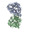
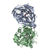
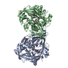
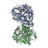
- PDB-7l0e: Crystal structure of bovine RPE65 in complex with gem-difluoro em... -
+ Open data

- Basic information
| Entry | Database: PDB / ID: 7l0e | ||||||
|---|---|---|---|---|---|---|---|
| Title | Crystal structure of bovine RPE65 in complex with gem-difluoro emixustat and palmitate | ||||||
 Components Components | Retinoid isomerohydrolase | ||||||
 Keywords Keywords | HYDROLASE/INHIBITOR / non-heme iron / beta propeller / isomerohydrolase / isomerase / monotopic membrane protein / HYDROLASE / HYDROLASE-INHIBITOR complex | ||||||
| Function / homology |  Function and homology information Function and homology informationretinoid isomerohydrolase / lutein isomerase / retinol isomerase activity / all-trans-retinyl-palmitate hydrolase, 11-cis retinol forming activity / all-trans-retinyl-ester hydrolase, 11-cis retinol forming activity / zeaxanthin biosynthetic process / beta-carotene 15,15'-dioxygenase activity / The canonical retinoid cycle in rods (twilight vision) / detection of light stimulus involved in visual perception / retinal metabolic process ...retinoid isomerohydrolase / lutein isomerase / retinol isomerase activity / all-trans-retinyl-palmitate hydrolase, 11-cis retinol forming activity / all-trans-retinyl-ester hydrolase, 11-cis retinol forming activity / zeaxanthin biosynthetic process / beta-carotene 15,15'-dioxygenase activity / The canonical retinoid cycle in rods (twilight vision) / detection of light stimulus involved in visual perception / retinal metabolic process / phosphatidylcholine binding / retina homeostasis / cardiolipin binding / phosphatidylserine binding / endoplasmic reticulum membrane / membrane / metal ion binding / identical protein binding / nucleus / plasma membrane Similarity search - Function | ||||||
| Biological species |  | ||||||
| Method |  X-RAY DIFFRACTION / SYNCHROTRON / FOURIER SYNTHESIS / Resolution: 1.9 Å X-RAY DIFFRACTION / SYNCHROTRON / FOURIER SYNTHESIS / Resolution: 1.9 Å | ||||||
 Authors Authors | Kiser, P.D. | ||||||
 Citation Citation | Journal: J.Med.Chem. / Year: 2021 Title: Rational Alteration of Pharmacokinetics of Chiral Fluorinated and Deuterated Derivatives of Emixustat for Retinal Therapy. Authors: Blum, E. / Zhang, J. / Zaluski, J. / Einstein, D.E. / Korshin, E.E. / Kubas, A. / Gruzman, A. / Tochtrop, G.P. / Kiser, P.D. / Palczewski, K. | ||||||
| History |
|
- Structure visualization
| Structure viewer | Molecule: MolmilJmol/JSmol |
|---|
- Downloads & links
-Download
| PDBx/mmCIF format | 7l0e.cif.gz | 240.1 KB | Display | PDBx/mmCIF format |
|---|---|---|---|---|
| PDB format | pdb7l0e.ent.gz | 189 KB | Display | PDB format |
| PDBx/mmJSON format | 7l0e.json.gz | Tree view | PDBx/mmJSON format | |
| Others |  Other downloads Other downloads |
-Validation report
| Arichive directory | https://data.pdbj.org/pub/pdb/validation_reports/l0/7l0eftp://data.pdbj.org/pub/pdb/validation_reports/l0/7l0e | HTTPS FTP |
|---|
-Related structure data
| Related structure data | 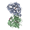 7k88C 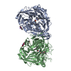 7k89C  7k8gC 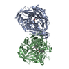 4rscS S: Starting model for refinement C: citing same article ( |
|---|---|
| Similar structure data |


-Links
 PDBj
PDBj


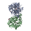
- Assembly
| Deposited unit | 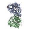
| ||||||||||||||||||
|---|---|---|---|---|---|---|---|---|---|---|---|---|---|---|---|---|---|---|---|
| 1 |
| ||||||||||||||||||
| Unit cell |
| ||||||||||||||||||
| Noncrystallographic symmetry (NCS) | NCS domain:
NCS domain segments: Component-ID: _ / Ens-ID: 1 / Beg auth comp-ID: SER / Beg label comp-ID: SER / End auth comp-ID: SER / End label comp-ID: SER / Refine code: _ / Auth seq-ID: 3 - 533 / Label seq-ID: 3 - 533
|
-Components
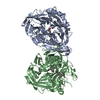
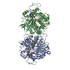
-Protein , 1 types, 2 molecules AB
| #1: Protein | Mass: 61040.195 Da / Num. of mol.: 2 Source method: isolated from a genetically manipulated source Source: (gene. exp.) References: UniProt: Q28175, retinoid isomerohydrolase, lutein isomerase |
|---|
-Non-polymers , 5 types, 851 molecules 

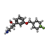


| #2: Chemical |  Mass: 55.845 Da / Num. of mol.: 2 / Source method: obtained synthetically / Formula: Fe / Feature type: SUBJECT OF INVESTIGATION Mass: 55.845 Da / Num. of mol.: 2 / Source method: obtained synthetically / Formula: Fe / Feature type: SUBJECT OF INVESTIGATION#3: Chemical |  Mass: 256.424 Da / Num. of mol.: 2 / Source method: obtained synthetically / Formula: C16H32O2 / Feature type: SUBJECT OF INVESTIGATION Mass: 256.424 Da / Num. of mol.: 2 / Source method: obtained synthetically / Formula: C16H32O2 / Feature type: SUBJECT OF INVESTIGATION#4: Chemical |  Mass: 299.356 Da / Num. of mol.: 2 / Source method: obtained synthetically / Formula: C16H23F2NO2 / Feature type: SUBJECT OF INVESTIGATION Mass: 299.356 Da / Num. of mol.: 2 / Source method: obtained synthetically / Formula: C16H23F2NO2 / Feature type: SUBJECT OF INVESTIGATION#5: Chemical | ChemComp-PO4 / |  Mass: 94.971 Da / Num. of mol.: 1 / Source method: obtained synthetically / Formula: PO4 Mass: 94.971 Da / Num. of mol.: 1 / Source method: obtained synthetically / Formula: PO4#6: Water | ChemComp-HOH / | Mass: 18.015 Da / Num. of mol.: 844 / Source method: isolated from a natural source / Formula: H2O |
|---|
-Details
| Has ligand of interest | Y |
|---|
-Experimental details
-Experiment
| Experiment | Method: X-RAY DIFFRACTION / Number of used crystals: 1 |
|---|
- Sample preparation
| Crystal | Density Matthews: 3.15 Å3/Da / Density % sol: 60.99 % |
|---|---|
| Crystal grow | Temperature: 281 K / Method: vapor diffusion, hanging drop Details: 30% PEG200, 100 mM Tris pH 8.5, 200 mM ammonium phosphate |
-Data collection
| Diffraction | Mean temperature: 100 K / Serial crystal experiment: N |
|---|---|
| Diffraction source | Source: SYNCHROTRON / Site: APS  / Beamline: 24-ID-E / Wavelength: 0.97918 Å / Beamline: 24-ID-E / Wavelength: 0.97918 Å |
| Detector | Type: DECTRIS EIGER X 16M / Detector: PIXEL / Date: Oct 16, 2020 |
| Radiation | Protocol: SINGLE WAVELENGTH / Monochromatic (M) / Laue (L): M / Scattering type: x-ray |
| Radiation wavelength | Wavelength: 0.97918 Å / Relative weight: 1 |
| Reflection | Resolution: 1.9→50 Å / Num. obs: 118471 / % possible obs: 99.7 % / Redundancy: 10.4 % / Biso Wilson estimate: 45 Å2 / CC1/2: 0.999 / Rmerge(I) obs: 0.113 / Net I/σ(I): 13.1 |
| Reflection shell | Resolution: 1.9→2.02 Å / Rmerge(I) obs: 2.482 / Mean I/σ(I) obs: 0.96 / Num. unique obs: 19003 / CC1/2: 0.43 |
- Processing
| Software |
| ||||||||||||||||||||||||||||||||||||||||||||||||||||||||||||
|---|---|---|---|---|---|---|---|---|---|---|---|---|---|---|---|---|---|---|---|---|---|---|---|---|---|---|---|---|---|---|---|---|---|---|---|---|---|---|---|---|---|---|---|---|---|---|---|---|---|---|---|---|---|---|---|---|---|---|---|---|---|
| Refinement | Method to determine structure: FOURIER SYNTHESIS Starting model: 4RSC Resolution: 1.9→47.87 Å / Cor.coef. Fo:Fc: 0.973 / Cor.coef. Fo:Fc free: 0.961 / SU B: 4.143 / SU ML: 0.107 / Cross valid method: THROUGHOUT / σ(F): 0 / ESU R: 0.12 / ESU R Free: 0.111 / Stereochemistry target values: MAXIMUM LIKELIHOOD Details: HYDROGENS HAVE BEEN ADDED IN THE RIDING POSITIONS U VALUES : REFINED INDIVIDUALLY
| ||||||||||||||||||||||||||||||||||||||||||||||||||||||||||||
| Solvent computation | Ion probe radii: 0.7 Å / Shrinkage radii: 0.7 Å / VDW probe radii: 1 Å / Solvent model: MASK | ||||||||||||||||||||||||||||||||||||||||||||||||||||||||||||
| Displacement parameters | Biso max: 127 Å2 / Biso mean: 44.77 Å2 / Biso min: 30.75 Å2
| ||||||||||||||||||||||||||||||||||||||||||||||||||||||||||||
| Refinement step | Cycle: final / Resolution: 1.9→47.87 Å
| ||||||||||||||||||||||||||||||||||||||||||||||||||||||||||||
| Refine LS restraints |
| ||||||||||||||||||||||||||||||||||||||||||||||||||||||||||||
| Refine LS restraints NCS | Ens-ID: 1 / Number: 16930 / Refine-ID: X-RAY DIFFRACTION / Type: interatomic distance / Rms dev position: 0.05 Å / Weight position: 0.05
| ||||||||||||||||||||||||||||||||||||||||||||||||||||||||||||
| LS refinement shell | Resolution: 1.904→1.953 Å / Rfactor Rfree error: 0 / Total num. of bins used: 20
|
