 Movie
Movie Controller


+ Open data

- Basic information
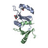
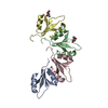
| Entry | Database: PDB / ID: 7kwy | ||||||
|---|---|---|---|---|---|---|---|
| Title | X-ray Crystal Structure of PlyCB Mutant R66K | ||||||
 Components Components | PlyCB | ||||||
 Keywords Keywords | ANTIMICROBIAL PROTEIN / ENDOLYSIN / VIRAL PROTEIN / PHAGE EFFECTOR PROTEIN | ||||||
| Function / homology | : / : / Streptococcus virus C1, PlyCB / killing of cells of another organism / identical protein binding / Endolysin PlyC, small cell-wall binding subunit Function and homology information Function and homology information | ||||||
| Biological species |  Streptococcus virus C1 Streptococcus virus C1 | ||||||
| Method |  X-RAY DIFFRACTION / SYNCHROTRON / MOLECULAR REPLACEMENT / Resolution: 1.7 Å X-RAY DIFFRACTION / SYNCHROTRON / MOLECULAR REPLACEMENT / Resolution: 1.7 Å | ||||||
 Authors Authors | Williams, D.E. / Broendum, S.S. / Hayes, B.K. / Drinkwater, N. / McGowan, S. | ||||||
 Citation Citation | Journal: Mol.Microbiol. / Year: 2021 Title: High avidity drives the interaction between the streptococcal C1 phage endolysin, PlyC, with the cell surface carbohydrates of Group A Streptococcus. Authors: Broendum, S.S. / Williams, D.E. / Hayes, B.K. / Kraus, F. / Fodor, J. / Clifton, B.E. / Geert Volbeda, A. / Codee, J.D.C. / Riley, B.T. / Drinkwater, N. / Farrow, K.A. / Tsyganov, K. / ...Authors: Broendum, S.S. / Williams, D.E. / Hayes, B.K. / Kraus, F. / Fodor, J. / Clifton, B.E. / Geert Volbeda, A. / Codee, J.D.C. / Riley, B.T. / Drinkwater, N. / Farrow, K.A. / Tsyganov, K. / Heselpoth, R.D. / Nelson, D.C. / Jackson, C.J. / Buckle, A.M. / McGowan, S. | ||||||
| History |
|
- Structure visualization
| Structure viewer | Molecule: MolmilJmol/JSmol |
|---|
- Downloads & links
-Download
| PDBx/mmCIF format | 7kwy.cif.gz | 72.2 KB | Display | PDBx/mmCIF format |
|---|---|---|---|---|
| PDB format | pdb7kwy.ent.gz | 50.7 KB | Display | PDB format |
| PDBx/mmJSON format | 7kwy.json.gz | Tree view | PDBx/mmJSON format | |
| Others |  Other downloads Other downloads |
-Validation report
| Arichive directory | https://data.pdbj.org/pub/pdb/validation_reports/kw/7kwyftp://data.pdbj.org/pub/pdb/validation_reports/kw/7kwy | HTTPS FTP |
|---|
-Related structure data
| Related structure data |  7kwtC  7kwwC  4f87S C: citing same article ( S: Starting model for refinement |
|---|---|
| Similar structure data |


-Links
 PDBj
PDBj- Assembly
| Deposited unit | 
| ||||||||||||
|---|---|---|---|---|---|---|---|---|---|---|---|---|---|
| 1 | 
| ||||||||||||
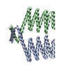
| Unit cell |
|
-Components
| #1: Protein | Mass: 7839.929 Da / Num. of mol.: 2 / Mutation: R66K Source method: isolated from a genetically manipulated source Source: (gene. exp.) Streptococcus virus C1 / Gene: orf9 / Production host:  #2: Chemical |   Mass: 118.174 Da / Num. of mol.: 2 / Source method: obtained synthetically / Formula: C6H14O2 / Comment: precipitant*YM Mass: 118.174 Da / Num. of mol.: 2 / Source method: obtained synthetically / Formula: C6H14O2 / Comment: precipitant*YM#3: Water | ChemComp-HOH / |  Mass: 18.015 Da / Num. of mol.: 114 / Source method: isolated from a natural source / Formula: H2O Mass: 18.015 Da / Num. of mol.: 114 / Source method: isolated from a natural source / Formula: H2OHas ligand of interest | N | |
|---|
-Experimental details
-Experiment
| Experiment | Method: X-RAY DIFFRACTION / Number of used crystals: 1 |
|---|
- Sample preparation
| Crystal | Density Matthews: 2.69 Å3/Da / Density % sol: 54.33 % |
|---|---|
| Crystal grow | Temperature: 293 K / Method: vapor diffusion / pH: 6 Details: 0.1M HEPES (pH 6.0), 0.2M sodium citrate, 25% methylpentanediol |
-Data collection
| Diffraction | Mean temperature: 100 K / Serial crystal experiment: N |
|---|---|
| Diffraction source | Source: SYNCHROTRON / Site: Australian Synchrotron  / Beamline: MX2 / Wavelength: 0.95366 Å / Beamline: MX2 / Wavelength: 0.95366 Å |
| Detector | Type: DECTRIS EIGER X 16M / Detector: PIXEL / Date: Mar 23, 2019 |
| Radiation | Protocol: SINGLE WAVELENGTH / Monochromatic (M) / Laue (L): M / Scattering type: x-ray |
| Radiation wavelength | Wavelength: 0.95366 Å / Relative weight: 1 |
| Reflection | Resolution: 1.7→37.85 Å / Num. obs: 21454 / % possible obs: 99.95 % / Redundancy: 2 % / Biso Wilson estimate: 29.02 Å2 / CC1/2: 0.987 / CC star: 0.997 / Net I/σ(I): 22.53 |
| Reflection shell | Resolution: 1.7→1.761 Å / Mean I/σ(I) obs: 1.65 / Num. unique obs: 2099 / CC1/2: 0.987 / CC star: 0.997 |
- Processing
| Software |
| |||||||||||||||||||||||||||||||||||||||||||||||||||||||||||||||
|---|---|---|---|---|---|---|---|---|---|---|---|---|---|---|---|---|---|---|---|---|---|---|---|---|---|---|---|---|---|---|---|---|---|---|---|---|---|---|---|---|---|---|---|---|---|---|---|---|---|---|---|---|---|---|---|---|---|---|---|---|---|---|---|---|
| Refinement | Method to determine structure: MOLECULAR REPLACEMENT Starting model: 4F87 Resolution: 1.7→37.85 Å / SU ML: 0.1695 / Cross valid method: FREE R-VALUE / σ(F): 1.35 / Phase error: 21.8089 Stereochemistry target values: GeoStd + Monomer Library + CDL v1.2
| |||||||||||||||||||||||||||||||||||||||||||||||||||||||||||||||
| Solvent computation | Shrinkage radii: 0.9 Å / VDW probe radii: 1.11 Å / Solvent model: FLAT BULK SOLVENT MODEL | |||||||||||||||||||||||||||||||||||||||||||||||||||||||||||||||
| Displacement parameters | Biso mean: 38.9 Å2 | |||||||||||||||||||||||||||||||||||||||||||||||||||||||||||||||
| Refinement step | Cycle: LAST / Resolution: 1.7→37.85 Å
| |||||||||||||||||||||||||||||||||||||||||||||||||||||||||||||||
| Refine LS restraints |
| |||||||||||||||||||||||||||||||||||||||||||||||||||||||||||||||
| LS refinement shell |
|
