 Movie
Movie Controller

[English] 日本語
 Yorodumi
- PDB-7kr6: Glycoside hydrolase family 16 endo-glucanase from Bacteroides ova... -
+ Open data

- Basic information
| Entry | Database: PDB / ID: 7kr6 | ||||||
|---|---|---|---|---|---|---|---|
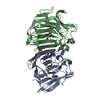
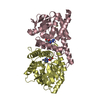
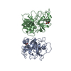
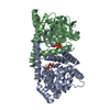
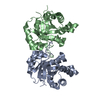
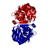
| Title | Glycoside hydrolase family 16 endo-glucanase from Bacteroides ovatus in complex with G4G3G-2F-DNP | ||||||
 Components Components | Glycoside hydrolase family 16 protein | ||||||
 Keywords Keywords | HYDROLASE / Inhibitor / glucanase | ||||||
| Function / homology | : / Glycosyl hydrolases family 16 / Glycoside hydrolase family 16 / Glycosyl hydrolases family 16 (GH16) domain profile. / hydrolase activity, hydrolyzing O-glycosyl compounds / Concanavalin A-like lectin/glucanase domain superfamily / carbohydrate metabolic process / beta-D-glucopyranose-(1-4)-beta-D-glucopyranose-(1-3)-2-deoxy-2-fluoro-alpha-D-glucopyranose / Glycoside hydrolase family 16 protein Function and homology information Function and homology information | ||||||
| Biological species |  Bacteroides ovatus (bacteria) Bacteroides ovatus (bacteria) | ||||||
| Method |  X-RAY DIFFRACTION / SYNCHROTRON / MOLECULAR REPLACEMENT / Resolution: 1.56 Å X-RAY DIFFRACTION / SYNCHROTRON / MOLECULAR REPLACEMENT / Resolution: 1.56 Å | ||||||
 Authors Authors | Tamura, K. / Brumer, H. / van Petegem, F. | ||||||
| Funding support |  Canada, 1items Canada, 1items
| ||||||
 Citation Citation | Journal: Acs Chem.Biol. / Year: 2021 Title: Orthogonal Active-Site Labels for Mixed-Linkage endo-beta-Glucanases. Authors: Jain, N. / Tamura, K. / Dejean, G. / Van Petegem, F. / Brumer, H. | ||||||
| History |
|
- Structure visualization
| Structure viewer | Molecule: MolmilJmol/JSmol |
|---|
- Downloads & links
-Download
| PDBx/mmCIF format | 7kr6.cif.gz | 193.8 KB | Display | PDBx/mmCIF format |
|---|---|---|---|---|
| PDB format | pdb7kr6.ent.gz | Display | PDB format | |
| PDBx/mmJSON format | 7kr6.json.gz | Tree view | PDBx/mmJSON format | |
| Others |  Other downloads Other downloads |
-Validation report
| Arichive directory | https://data.pdbj.org/pub/pdb/validation_reports/kr/7kr6ftp://data.pdbj.org/pub/pdb/validation_reports/kr/7kr6 | HTTPS FTP |
|---|
-Related structure data
| Related structure data | 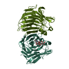 6vhoC  5nboS C: citing same article ( S: Starting model for refinement |
|---|---|
| Similar structure data |


-Links
 PDBj
PDBj
- Assembly
| Deposited unit | 
| ||||||||
|---|---|---|---|---|---|---|---|---|---|
| 1 |
| ||||||||
| Unit cell |
|
-Components
| #1: Protein | Mass: 30593.943 Da / Num. of mol.: 2 / Mutation: E148A Source method: isolated from a genetically manipulated source Source: (gene. exp.) Bacteroides ovatus (bacteria)Gene: DW206_07605, DW890_00285, F3D51_00680, F3D58_00285, F3F38_18210 Production host: #2: Polysaccharide | 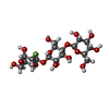  Source method: isolated from a genetically manipulated source References: beta-D-glucopyranose-(1-4)-beta-D-glucopyranose-(1-3)-2-deoxy-2-fluoro-alpha-D-glucopyranose #3: Water | ChemComp-HOH / |  Mass: 18.015 Da / Num. of mol.: 374 / Source method: isolated from a natural source / Formula: H2O Mass: 18.015 Da / Num. of mol.: 374 / Source method: isolated from a natural source / Formula: H2OHas ligand of interest | Y | |
|---|
-Experimental details
-Experiment
| Experiment | Method: X-RAY DIFFRACTION / Number of used crystals: 1 |
|---|
- Sample preparation
| Crystal | Density Matthews: 2.15 Å3/Da / Density % sol: 42.76 % |
|---|---|
| Crystal grow | Temperature: 278 K / Method: vapor diffusion, hanging drop Details: 0.2 M sodium chloride, 0.1 M Tris pH 8.5, 25% (w/v) PEG3350 |
-Data collection
| Diffraction | Mean temperature: 100 K / Serial crystal experiment: N |
|---|---|
| Diffraction source | Source: SYNCHROTRON / Site: SSRL  / Beamline: BL12-2 / Wavelength: 0.979 Å / Beamline: BL12-2 / Wavelength: 0.979 Å |
| Detector | Type: DECTRIS PILATUS3 S 6M / Detector: PIXEL / Date: Nov 7, 2020 |
| Radiation | Protocol: SINGLE WAVELENGTH / Monochromatic (M) / Laue (L): M / Scattering type: x-ray |
| Radiation wavelength | Wavelength: 0.979 Å / Relative weight: 1 |
| Reflection | Resolution: 1.56→39.4 Å / Num. obs: 143215 / % possible obs: 98.7 % / Redundancy: 2 % / CC1/2: 1 / Net I/σ(I): 14.2 |
| Reflection shell | Resolution: 1.56→1.59 Å / Num. unique obs: 6822 / CC1/2: 0.935 |
- Processing
| Software |
| ||||||||||||||||||||||||||||||||||||||||||||||||||||||||||||||||||||||||||||||||||||||||||||||||||||||||||||||||||||||||||||||||||||||||||||||||||||||
|---|---|---|---|---|---|---|---|---|---|---|---|---|---|---|---|---|---|---|---|---|---|---|---|---|---|---|---|---|---|---|---|---|---|---|---|---|---|---|---|---|---|---|---|---|---|---|---|---|---|---|---|---|---|---|---|---|---|---|---|---|---|---|---|---|---|---|---|---|---|---|---|---|---|---|---|---|---|---|---|---|---|---|---|---|---|---|---|---|---|---|---|---|---|---|---|---|---|---|---|---|---|---|---|---|---|---|---|---|---|---|---|---|---|---|---|---|---|---|---|---|---|---|---|---|---|---|---|---|---|---|---|---|---|---|---|---|---|---|---|---|---|---|---|---|---|---|---|---|---|---|---|
| Refinement | Method to determine structure: MOLECULAR REPLACEMENT Starting model: 5NBO Resolution: 1.56→39.4 Å / Cor.coef. Fo:Fc: 0.973 / Cor.coef. Fo:Fc free: 0.964 / Cross valid method: FREE R-VALUE / ESU R: 0.078 / ESU R Free: 0.078 Details: Hydrogens have been added in their riding positions
| ||||||||||||||||||||||||||||||||||||||||||||||||||||||||||||||||||||||||||||||||||||||||||||||||||||||||||||||||||||||||||||||||||||||||||||||||||||||
| Solvent computation | Ion probe radii: 0.8 Å / Shrinkage radii: 0.8 Å / VDW probe radii: 1.2 Å / Solvent model: MASK BULK SOLVENT | ||||||||||||||||||||||||||||||||||||||||||||||||||||||||||||||||||||||||||||||||||||||||||||||||||||||||||||||||||||||||||||||||||||||||||||||||||||||
| Displacement parameters | Biso mean: 24.81 Å2
| ||||||||||||||||||||||||||||||||||||||||||||||||||||||||||||||||||||||||||||||||||||||||||||||||||||||||||||||||||||||||||||||||||||||||||||||||||||||
| Refinement step | Cycle: LAST / Resolution: 1.56→39.4 Å
| ||||||||||||||||||||||||||||||||||||||||||||||||||||||||||||||||||||||||||||||||||||||||||||||||||||||||||||||||||||||||||||||||||||||||||||||||||||||
| Refine LS restraints |
| ||||||||||||||||||||||||||||||||||||||||||||||||||||||||||||||||||||||||||||||||||||||||||||||||||||||||||||||||||||||||||||||||||||||||||||||||||||||
| LS refinement shell |
|
