 Movie
Movie Controller


+ Open data

- Basic information
| Entry | Database: PDB / ID: 7kkb | ||||||
|---|---|---|---|---|---|---|---|
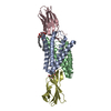
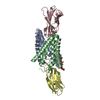
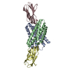
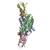
| Title | Fluoride channel Fluc-Ec2 mutant S81C with bromide | ||||||
 Components Components |
| ||||||
 Keywords Keywords | MEMBRANE PROTEIN / fluoride channel | ||||||
| Function / homology |  Function and homology information Function and homology informationfluoride channel activity / cellular detoxification of fluoride / metal ion binding / plasma membrane Similarity search - Function | ||||||
| Biological species |   Homo sapiens (human) Homo sapiens (human) | ||||||
| Method |  X-RAY DIFFRACTION / SYNCHROTRON / MOLECULAR REPLACEMENT / Resolution: 2.9 Å X-RAY DIFFRACTION / SYNCHROTRON / MOLECULAR REPLACEMENT / Resolution: 2.9 Å | ||||||
 Authors Authors | McIlwain, B.C. / Stockbridge, R.B. | ||||||
| Funding support |  United States, 1items United States, 1items
| ||||||
 Citation Citation | Journal: Elife / Year: 2021 Title: The fluoride permeation pathway and anion recognition in Fluc family fluoride channels. Authors: McIlwain, B.C. / Gundepudi, R. / Koff, B.B. / Stockbridge, R.B. | ||||||
| History |
|
- Structure visualization
| Structure viewer | Molecule: MolmilJmol/JSmol |
|---|
- Downloads & links
-Download
| PDBx/mmCIF format | 7kkb.cif.gz | 188.1 KB | Display | PDBx/mmCIF format |
|---|---|---|---|---|
| PDB format | pdb7kkb.ent.gz | 148.9 KB | Display | PDB format |
| PDBx/mmJSON format | 7kkb.json.gz | Tree view | PDBx/mmJSON format | |
| Others |  Other downloads Other downloads |
-Validation report
| Summary document | 7kkb_validation.pdf.gz | 998.7 KB | Display | wwPDB validaton report |
|---|---|---|---|---|
| Full document | 7kkb_full_validation.pdf.gz | 998.7 KB | Display | |
| Data in XML | 7kkb_validation.xml.gz | 16.8 KB | Display | |
| Data in CIF | 7kkb_validation.cif.gz | 22.3 KB | Display | |
| Arichive directory | https://data.pdbj.org/pub/pdb/validation_reports/kk/7kkbftp://data.pdbj.org/pub/pdb/validation_reports/kk/7kkb | HTTPS FTP |
-Related structure data
| Related structure data |  7kk8C  7kk9C  7kkaC  7kkrC  5a43S S: Starting model for refinement C: citing same article ( |
|---|---|
| Similar structure data |


-Links
 PDBj
PDBj


- Assembly
| Deposited unit | 
| ||||||||
|---|---|---|---|---|---|---|---|---|---|
| 1 |
| ||||||||
| Unit cell |
|
-Components
-Protein / Antibody / Sugars , 3 types, 7 molecules ABCD
| #1: Protein | Mass: 13599.307 Da / Num. of mol.: 2 / Mutation: S81C Source method: isolated from a genetically manipulated source Source: (gene. exp.) #2: Antibody | Mass: 10375.529 Da / Num. of mol.: 2 Source method: isolated from a genetically manipulated source Source: (gene. exp.) Homo sapiens (human) / Production host: #6: Sugar |  Type: D-saccharide / Mass: 482.562 Da / Num. of mol.: 3 / Source method: obtained synthetically / Formula: C22H42O11 / Comment: detergent*YM Type: D-saccharide / Mass: 482.562 Da / Num. of mol.: 3 / Source method: obtained synthetically / Formula: C22H42O11 / Comment: detergent*YM |
|---|
-Non-polymers , 4 types, 7 molecules 


| #3: Chemical |  Mass: 79.904 Da / Num. of mol.: 2 / Source method: obtained synthetically / Formula: Br Mass: 79.904 Da / Num. of mol.: 2 / Source method: obtained synthetically / Formula: Br#4: Chemical | ChemComp-NA / |  Mass: 22.990 Da / Num. of mol.: 1 / Source method: obtained synthetically / Formula: Na Mass: 22.990 Da / Num. of mol.: 1 / Source method: obtained synthetically / Formula: Na#5: Chemical |  Mass: 18.998 Da / Num. of mol.: 2 / Source method: obtained synthetically / Formula: F Mass: 18.998 Da / Num. of mol.: 2 / Source method: obtained synthetically / Formula: F#7: Water | ChemComp-HOH / | Mass: 18.015 Da / Num. of mol.: 2 / Source method: isolated from a natural source / Formula: H2O |
|---|
-Details
| Has ligand of interest | N |
|---|
-Experimental details
-Experiment
| Experiment | Method: X-RAY DIFFRACTION / Number of used crystals: 1 |
|---|
- Sample preparation
| Crystal | Density Matthews: 5.68 Å3/Da / Density % sol: 78.34 % |
|---|---|
| Crystal grow | Temperature: 298 K / Method: vapor diffusion / Details: 0.1M glycine pH 9 36% PEG 600 |
-Data collection
| Diffraction | Mean temperature: 80 K / Serial crystal experiment: N | ||||||||||||||||||||||||||||||
|---|---|---|---|---|---|---|---|---|---|---|---|---|---|---|---|---|---|---|---|---|---|---|---|---|---|---|---|---|---|---|---|
| Diffraction source | Source: SYNCHROTRON / Site: APS / Beamline: 21-ID-D / Wavelength: 0.91836 Å | ||||||||||||||||||||||||||||||
| Detector | Type: DECTRIS EIGER2 X 9M / Detector: PIXEL / Date: Feb 28, 2018 | ||||||||||||||||||||||||||||||
| Radiation | Protocol: SINGLE WAVELENGTH / Monochromatic (M) / Laue (L): M / Scattering type: x-ray | ||||||||||||||||||||||||||||||
| Radiation wavelength | Wavelength: 0.91836 Å / Relative weight: 1 | ||||||||||||||||||||||||||||||
| Reflection | Resolution: 2.9→46.65 Å / Num. obs: 23623 / % possible obs: 99.9 % / Redundancy: 12.5 % / CC1/2: 0.981 / Rmerge(I) obs: 0.37 / Rpim(I) all: 0.108 / Rrim(I) all: 0.386 / Net I/σ(I): 9.8 / Num. measured all: 295813 / Scaling rejects: 112 | ||||||||||||||||||||||||||||||
| Reflection shell | Diffraction-ID: 1
|
- Processing
| Software |
| ||||||||||||||||||||||||||||||||||||||||||||||||||||||||||||||||||
|---|---|---|---|---|---|---|---|---|---|---|---|---|---|---|---|---|---|---|---|---|---|---|---|---|---|---|---|---|---|---|---|---|---|---|---|---|---|---|---|---|---|---|---|---|---|---|---|---|---|---|---|---|---|---|---|---|---|---|---|---|---|---|---|---|---|---|---|
| Refinement | Method to determine structure: MOLECULAR REPLACEMENT Starting model: 5A43 Resolution: 2.9→46.65 Å / SU ML: 0.4 / Cross valid method: THROUGHOUT / σ(F): 1.35 / Phase error: 29.18 / Stereochemistry target values: ML
| ||||||||||||||||||||||||||||||||||||||||||||||||||||||||||||||||||
| Solvent computation | Shrinkage radii: 0.9 Å / VDW probe radii: 1.11 Å / Solvent model: FLAT BULK SOLVENT MODEL | ||||||||||||||||||||||||||||||||||||||||||||||||||||||||||||||||||
| Displacement parameters | Biso max: 211.3 Å2 / Biso mean: 68.6302 Å2 / Biso min: 33.55 Å2 | ||||||||||||||||||||||||||||||||||||||||||||||||||||||||||||||||||
| Refinement step | Cycle: final / Resolution: 2.9→46.65 Å
| ||||||||||||||||||||||||||||||||||||||||||||||||||||||||||||||||||
| LS refinement shell | Refine-ID: X-RAY DIFFRACTION / Rfactor Rfree error: 0 / Total num. of bins used: 8 / % reflection obs: 100 %
| ||||||||||||||||||||||||||||||||||||||||||||||||||||||||||||||||||
| Refinement TLS params. | Method: refined / Origin x: 1.527 Å / Origin y: -42.7316 Å / Origin z: -21.6967 Å
| ||||||||||||||||||||||||||||||||||||||||||||||||||||||||||||||||||
| Refinement TLS group |
|
