 Movie
Movie Controller

[English] 日本語
 Yorodumi
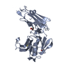
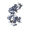
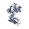
- PDB-7kem: Crystallographic structure of L,D-transpeptidase 2 from Mycobacte... -
+ Open data

- Basic information
| Entry | Database: PDB / ID: 7kem | ||||||
|---|---|---|---|---|---|---|---|
| Title | Crystallographic structure of L,D-transpeptidase 2 from Mycobacterium tuberculosis | ||||||
 Components Components | L,D-transpeptidase 2 | ||||||
 Keywords Keywords | TRANSFERASE / Transpeptidase / Peptidoglycan synthesis | ||||||
| Function / homology |  Function and homology information Function and homology informationpeptidoglycan-protein cross-linking / peptidoglycan L,D-transpeptidase activity / Transferases; Acyltransferases; Aminoacyltransferases / acyltransferase activity / cell wall organization / regulation of cell shape / metal ion binding / plasma membrane Similarity search - Function | ||||||
| Biological species |   Mycobacterium tuberculosis (bacteria) Mycobacterium tuberculosis (bacteria) | ||||||
| Method |  X-RAY DIFFRACTION / SYNCHROTRON / MOLECULAR REPLACEMENT / Resolution: 1.77 Å X-RAY DIFFRACTION / SYNCHROTRON / MOLECULAR REPLACEMENT / Resolution: 1.77 Å | ||||||
 Authors Authors | Libreros, G.A. / Dias, M.V.B. | ||||||
| Funding support |  Portugal, 1items Portugal, 1items
| ||||||
 Citation Citation | Journal: To Be Published Title: Crystallographic structure of L,D-transpeptidase 2 from Mycobacterium tuberculosis. Authors: Libreros, G.A. / Dias, M.V.B. | ||||||
| History |
|
- Structure visualization
| Structure viewer | Molecule: MolmilJmol/JSmol |
|---|
- Downloads & links
-Download
| PDBx/mmCIF format | 7kem.cif.gz | 120.8 KB | Display | PDBx/mmCIF format |
|---|---|---|---|---|
| PDB format | pdb7kem.ent.gz | 91.1 KB | Display | PDB format |
| PDBx/mmJSON format | 7kem.json.gz | Tree view | PDBx/mmJSON format | |
| Others |  Other downloads Other downloads |
-Validation report
| Arichive directory | https://data.pdbj.org/pub/pdb/validation_reports/ke/7kemftp://data.pdbj.org/pub/pdb/validation_reports/ke/7kem | HTTPS FTP |
|---|
-Related structure data
| Related structure data |  3turS S: Starting model for refinement |
|---|---|
| Similar structure data |


-Links
 PDBj
PDBj


- Assembly
| Deposited unit | 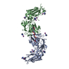
| ||||||||
|---|---|---|---|---|---|---|---|---|---|
| 1 | 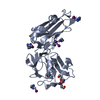
| ||||||||
| 2 | 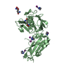
| ||||||||
| Unit cell |
|
-Components
| #1: Protein | Mass: 43407.711 Da / Num. of mol.: 2 Source method: isolated from a genetically manipulated source Source: (gene. exp.) Mycobacterium tuberculosis (strain ATCC 25618 / H37Rv) (bacteria)Strain: ATCC 25618 / H37Rv / Gene: ldtB, lppS, Rv2518c, RVBD_2518c, P425_02624 / Plasmid: pET28a / Production host: References: UniProt: I6Y9J2, Transferases; Acyltransferases; Aminoacyltransferases #2: Chemical | ChemComp-0JC /   Mass: 764.162 Da / Num. of mol.: 8 / Source method: obtained synthetically / Formula: C4H16I2N4Pt2 Mass: 764.162 Da / Num. of mol.: 8 / Source method: obtained synthetically / Formula: C4H16I2N4Pt2#3: Chemical | ChemComp-DGL / | 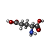  Type: D-peptide linking / Mass: 147.129 Da / Num. of mol.: 1 / Source method: obtained synthetically / Formula: C5H9NO4 Type: D-peptide linking / Mass: 147.129 Da / Num. of mol.: 1 / Source method: obtained synthetically / Formula: C5H9NO4#4: Chemical | ChemComp-6CL / |   Type: L-peptide linking / Mass: 191.205 Da / Num. of mol.: 1 / Source method: obtained synthetically / Formula: C7H15N2O4 Type: L-peptide linking / Mass: 191.205 Da / Num. of mol.: 1 / Source method: obtained synthetically / Formula: C7H15N2O4#5: Chemical | ChemComp-PT / |   Mass: 195.078 Da / Num. of mol.: 1 / Source method: obtained synthetically / Formula: Pt Mass: 195.078 Da / Num. of mol.: 1 / Source method: obtained synthetically / Formula: PtHas ligand of interest | N | |
|---|
-Experimental details
-Experiment
| Experiment | Method: X-RAY DIFFRACTION / Number of used crystals: 1 |
|---|
- Sample preparation
| Crystal | Density Matthews: 2.51 Å3/Da / Density % sol: 51.06 % |
|---|---|
| Crystal grow | Temperature: 293 K / Method: vapor diffusion, hanging drop / pH: 6 / Details: 3M NaOAc |
-Data collection
| Diffraction | Mean temperature: 100 K / Serial crystal experiment: N |
|---|---|
| Diffraction source | Source: SYNCHROTRON / Site: LNLS  / Beamline: W01B-MX2 / Wavelength: 1.458 Å / Beamline: W01B-MX2 / Wavelength: 1.458 Å |
| Detector | Type: DECTRIS PILATUS 2M / Detector: PIXEL / Date: Jul 15, 2015 |
| Radiation | Protocol: SINGLE WAVELENGTH / Monochromatic (M) / Laue (L): M / Scattering type: x-ray |
| Radiation wavelength | Wavelength: 1.458 Å / Relative weight: 1 |
| Reflection | Resolution: 1.77→49.66 Å / Num. obs: 80670 / % possible obs: 99.6 % / Redundancy: 12.3 % / Biso Wilson estimate: 22.8 Å2 / CC1/2: 0.999 / Rmerge(I) obs: 0.099 / Rrim(I) all: 0.108 / Net I/σ(I): 15.6 |
| Reflection shell | Resolution: 1.77→1.83 Å / Redundancy: 9.9 % / Rmerge(I) obs: 0.981 / Mean I/σ(I) obs: 2.2 / Num. unique obs: 8453 / CC1/2: 0.824 / % possible all: 99.9 |
- Processing
| Software |
| |||||||||||||||||||||||||||||||||||||||||||||
|---|---|---|---|---|---|---|---|---|---|---|---|---|---|---|---|---|---|---|---|---|---|---|---|---|---|---|---|---|---|---|---|---|---|---|---|---|---|---|---|---|---|---|---|---|---|---|
| Refinement | Method to determine structure: MOLECULAR REPLACEMENT Starting model: 3TUR Resolution: 1.77→49.66 Å / Cor.coef. Fo:Fc: 0.923 / Cor.coef. Fo:Fc free: 0.914 / SU B: 2.876 / SU ML: 0.089 / Cross valid method: THROUGHOUT / σ(F): 0 / ESU R: 0.118 / ESU R Free: 0.111 / Stereochemistry target values: MAXIMUM LIKELIHOOD / Details: U VALUES : REFINED INDIVIDUALLY
| |||||||||||||||||||||||||||||||||||||||||||||
| Solvent computation | Ion probe radii: 0.8 Å / Shrinkage radii: 0.8 Å / VDW probe radii: 1.2 Å / Solvent model: MASK | |||||||||||||||||||||||||||||||||||||||||||||
| Displacement parameters | Biso max: 94.58 Å2 / Biso mean: 25.543 Å2 / Biso min: 13.35 Å2
| |||||||||||||||||||||||||||||||||||||||||||||
| Refinement step | Cycle: final / Resolution: 1.77→49.66 Å
| |||||||||||||||||||||||||||||||||||||||||||||
| Refine LS restraints |
| |||||||||||||||||||||||||||||||||||||||||||||
| LS refinement shell | Resolution: 1.77→1.816 Å / Rfactor Rfree error: 0 / Total num. of bins used: 20
|
