 Movie
Movie Controller

[English] 日本語
 Yorodumi
- PDB-7kc7: Biotin Carboxylase domain of Thermophilic 2-Oxoglutarate Carboxyl... -
+ Open data

- Basic information
| Entry | Database: PDB / ID: 7kc7 | ||||||
|---|---|---|---|---|---|---|---|
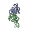
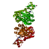
| Title | Biotin Carboxylase domain of Thermophilic 2-Oxoglutarate Carboxylase bound to ADP without Magnesium with disordered phosphate tail | ||||||
 Components Components | 2-oxoglutarate carboxylase small subunit | ||||||
 Keywords Keywords | LIGASE / biotin carboxylase / biotin-dependent carboxylase / pyruvate carboxylase / ATP-grasp / Aquificales / rTCA / dimer interface / bicarbonate / Sequence Determining Positions / structural waters / thermophile / carbon fixation / thermophilic protein / dimer / wet interface | ||||||
| Function / homology |  Function and homology information Function and homology information2-oxoglutarate carboxylase / 2-oxoglutarate carboxylase activity / ATP binding / metal ion binding Similarity search - Function | ||||||
| Biological species |   Hydrogenobacter thermophilus (bacteria) Hydrogenobacter thermophilus (bacteria) | ||||||
| Method |  X-RAY DIFFRACTION / SYNCHROTRON / MOLECULAR REPLACEMENT / Resolution: 2.2 Å X-RAY DIFFRACTION / SYNCHROTRON / MOLECULAR REPLACEMENT / Resolution: 2.2 Å | ||||||
| Model details | Nucleotide free dimer with Open Lid domain | ||||||
 Authors Authors | Buhrman, G.K. / Rose, R.B. / Enriquez, P. / Truong, V. | ||||||
| Funding support |  United States, 1items United States, 1items
| ||||||
 Citation Citation | Journal: Biochemistry / Year: 2021 Title: Structure, Function, and Thermal Adaptation of the Biotin Carboxylase Domain Dimer from Hydrogenobacter thermophilus 2-Oxoglutarate Carboxylase. Authors: Buhrman, G. / Enriquez, P. / Dillard, L. / Baer, H. / Truong, V. / Grunden, A.M. / Rose, R.B. | ||||||
| History |
|
- Structure visualization
| Structure viewer | Molecule: MolmilJmol/JSmol |
|---|
- Downloads & links
-Download
| PDBx/mmCIF format | 7kc7.cif.gz | 335.6 KB | Display | PDBx/mmCIF format |
|---|---|---|---|---|
| PDB format | pdb7kc7.ent.gz | 272.3 KB | Display | PDB format |
| PDBx/mmJSON format | 7kc7.json.gz | Tree view | PDBx/mmJSON format | |
| Others |  Other downloads Other downloads |
-Validation report
| Arichive directory | https://data.pdbj.org/pub/pdb/validation_reports/kc/7kc7ftp://data.pdbj.org/pub/pdb/validation_reports/kc/7kc7 | HTTPS FTP |
|---|
-Related structure data
| Related structure data | 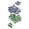 7kblC 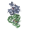 7kctC 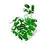 1ulzS S: Starting model for refinement C: citing same article ( |
|---|---|
| Similar structure data |


-Links
 PDBj
PDBj


- Assembly
| Deposited unit | 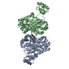
| ||||||||
|---|---|---|---|---|---|---|---|---|---|
| 1 |
| ||||||||
| Unit cell |
|
-Components
| #1: Protein | Mass: 55014.836 Da / Num. of mol.: 2 / Fragment: Biotin Carboxylase Domain Source method: isolated from a genetically manipulated source Source: (gene. exp.) Hydrogenobacter thermophilus (bacteria)Gene: cfiB, HTH_1393, Hydth_1383 / Plasmid: pET24b / Production host: #2: Chemical |   Mass: 94.971 Da / Num. of mol.: 2 / Source method: obtained synthetically / Formula: PO4 Mass: 94.971 Da / Num. of mol.: 2 / Source method: obtained synthetically / Formula: PO4#3: Chemical | ChemComp-ADP / |   Mass: 427.201 Da / Num. of mol.: 1 / Source method: obtained synthetically / Formula: C10H15N5O10P2 / Feature type: SUBJECT OF INVESTIGATION / Comment: ADP, energy-carrying molecule*YM Mass: 427.201 Da / Num. of mol.: 1 / Source method: obtained synthetically / Formula: C10H15N5O10P2 / Feature type: SUBJECT OF INVESTIGATION / Comment: ADP, energy-carrying molecule*YM#4: Water | ChemComp-HOH / |  Mass: 18.015 Da / Num. of mol.: 110 / Source method: isolated from a natural source / Formula: H2O Mass: 18.015 Da / Num. of mol.: 110 / Source method: isolated from a natural source / Formula: H2OHas ligand of interest | Y | |
|---|
-Experimental details
-Experiment
| Experiment | Method: X-RAY DIFFRACTION / Number of used crystals: 1 |
|---|
- Sample preparation
| Crystal | Density Matthews: 2.21 Å3/Da / Density % sol: 44.25 % |
|---|---|
| Crystal grow | Temperature: 291 K / Method: vapor diffusion, hanging drop / pH: 8 Details: PEG 3350, 0.02M Citric Acid, 0.08M Bis-Tris-Propane |
-Data collection
| Diffraction | Mean temperature: 100 K / Serial crystal experiment: N |
|---|---|
| Diffraction source | Source: SYNCHROTRON / Site: APS / Beamline: 22-ID / Wavelength: 1 Å |
| Detector | Type: MARMOSAIC 300 mm CCD / Detector: CCD / Date: Dec 5, 2013 |
| Radiation | Protocol: SINGLE WAVELENGTH / Monochromatic (M) / Laue (L): M / Scattering type: x-ray |
| Radiation wavelength | Wavelength: 1 Å / Relative weight: 1 |
| Reflection | Resolution: 2.2→50 Å / Num. obs: 50075 / % possible obs: 99.6 % / Redundancy: 6.8 % / Biso Wilson estimate: 40.77 Å2 / Rsym value: 0.112 / Net I/σ(I): 26.54 |
| Reflection shell | Resolution: 2.2→2.24 Å / Redundancy: 5.9 % / Num. unique obs: 2489 / Rsym value: 1.29 / % possible all: 100 |
- Processing
| Software |
| |||||||||||||||||||||||||||||||||||||||||||||||||||||||||||||||||||||||||||||||||||||||||||||||||||||||||
|---|---|---|---|---|---|---|---|---|---|---|---|---|---|---|---|---|---|---|---|---|---|---|---|---|---|---|---|---|---|---|---|---|---|---|---|---|---|---|---|---|---|---|---|---|---|---|---|---|---|---|---|---|---|---|---|---|---|---|---|---|---|---|---|---|---|---|---|---|---|---|---|---|---|---|---|---|---|---|---|---|---|---|---|---|---|---|---|---|---|---|---|---|---|---|---|---|---|---|---|---|---|---|---|---|---|---|
| Refinement | Method to determine structure: MOLECULAR REPLACEMENT Starting model: pdbid 1ULZ Resolution: 2.2→44.17 Å / SU ML: 0.23 / Cross valid method: THROUGHOUT / σ(F): 1.36 / Phase error: 24.35 / Stereochemistry target values: ML
| |||||||||||||||||||||||||||||||||||||||||||||||||||||||||||||||||||||||||||||||||||||||||||||||||||||||||
| Solvent computation | Shrinkage radii: 0.9 Å / VDW probe radii: 1.11 Å / Solvent model: FLAT BULK SOLVENT MODEL | |||||||||||||||||||||||||||||||||||||||||||||||||||||||||||||||||||||||||||||||||||||||||||||||||||||||||
| Displacement parameters | Biso max: 206.59 Å2 / Biso mean: 52.3385 Å2 / Biso min: 24.18 Å2 | |||||||||||||||||||||||||||||||||||||||||||||||||||||||||||||||||||||||||||||||||||||||||||||||||||||||||
| Refinement step | Cycle: final / Resolution: 2.2→44.17 Å
| |||||||||||||||||||||||||||||||||||||||||||||||||||||||||||||||||||||||||||||||||||||||||||||||||||||||||
| LS refinement shell | Refine-ID: X-RAY DIFFRACTION / Rfactor Rfree error: 0 / Total num. of bins used: 14
|
