 Movie
Movie Controller

[English] 日本語
 Yorodumi
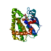
- PDB-7k7v: The X-ray crystal structure of SSR4, an S. pombe chromatin remode... -
+ Open data

- Basic information
| Entry | Database: PDB / ID: 7k7v | ||||||
|---|---|---|---|---|---|---|---|
| Title | The X-ray crystal structure of SSR4, an S. pombe chromatin remodelling protein: iodide derivative | ||||||
 Components Components | SWI/SNF and RSC complexes subunit ssr4 | ||||||
 Keywords Keywords | GENE REGULATION / chromatin remodelling / SAD phasing / novel structure | ||||||
| Function / homology |  Function and homology information Function and homology informationRSC-type complex / SWI/SNF complex / transcription initiation-coupled chromatin remodeling / chromatin remodeling / regulation of transcription by RNA polymerase II / chromatin / nucleus / cytosol Similarity search - Function | ||||||
| Biological species |  | ||||||
| Method |  X-RAY DIFFRACTION / SYNCHROTRON / SAD / Resolution: 1.882 Å X-RAY DIFFRACTION / SYNCHROTRON / SAD / Resolution: 1.882 Å | ||||||
 Authors Authors | Peat, T.S. / Newman, J. | ||||||
 Citation Citation | Journal: Acta Crystallogr.,Sect.F / Year: 2020 Title: The X-ray crystal structure of the N-terminal domain of Ssr4, a Schizosaccharomyces pombe chromatin-remodelling protein. Authors: Newman, J. / Nebl, T. / Van, H. / Peat, T.S. | ||||||
| History |
|
- Structure visualization
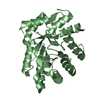
| Structure viewer | Molecule: MolmilJmol/JSmol |
|---|
- Downloads & links
-Download
| PDBx/mmCIF format | 7k7v.cif.gz | 57.5 KB | Display | PDBx/mmCIF format |
|---|---|---|---|---|
| PDB format | pdb7k7v.ent.gz | 38.3 KB | Display | PDB format |
| PDBx/mmJSON format | 7k7v.json.gz | Tree view | PDBx/mmJSON format | |
| Others |  Other downloads Other downloads |
-Validation report
| Arichive directory | https://data.pdbj.org/pub/pdb/validation_reports/k7/7k7vftp://data.pdbj.org/pub/pdb/validation_reports/k7/7k7v | HTTPS FTP |
|---|
-Related structure data


-Links
 PDBj
PDBj- Assembly
| Deposited unit | 
| ||||||||
|---|---|---|---|---|---|---|---|---|---|
| 1 |
| ||||||||
| Unit cell |
|
-Components
| #1: Protein | Mass: 22334.494 Da / Num. of mol.: 1 Source method: isolated from a genetically manipulated source Source: (gene. exp.) Gene: ssr4, SPBP23A10.05 / Production host:  | ||||||
|---|---|---|---|---|---|---|---|
| #2: Chemical | ChemComp-GOL /   Mass: 92.094 Da / Num. of mol.: 1 / Source method: obtained synthetically / Formula: C3H8O3 Mass: 92.094 Da / Num. of mol.: 1 / Source method: obtained synthetically / Formula: C3H8O3 | ||||||
| #3: Chemical | ChemComp-CL /   Mass: 35.453 Da / Num. of mol.: 4 / Source method: obtained synthetically / Formula: Cl Mass: 35.453 Da / Num. of mol.: 4 / Source method: obtained synthetically / Formula: Cl#4: Chemical |   Mass: 126.904 Da / Num. of mol.: 2 / Source method: obtained synthetically / Formula: I Mass: 126.904 Da / Num. of mol.: 2 / Source method: obtained synthetically / Formula: I#5: Water | ChemComp-HOH / |  Mass: 18.015 Da / Num. of mol.: 89 / Source method: isolated from a natural source / Formula: H2O Mass: 18.015 Da / Num. of mol.: 89 / Source method: isolated from a natural source / Formula: H2OHas ligand of interest | N | |
-Experimental details
-Experiment
| Experiment | Method: X-RAY DIFFRACTION / Number of used crystals: 1 |
|---|
- Sample preparation
| Crystal | Density Matthews: 2.56 Å3/Da / Density % sol: 51.94 % |
|---|---|
| Crystal grow | Temperature: 281 K / Method: vapor diffusion, sitting drop Details: Crystallisation experiments were set up in SD2 sitting drop plates at 8 C with 200 nL protein plus 200 nL reservoir with 50 uL of reservoir in the wells. The protein concentration was 5 ...Details: Crystallisation experiments were set up in SD2 sitting drop plates at 8 C with 200 nL protein plus 200 nL reservoir with 50 uL of reservoir in the wells. The protein concentration was 5 mg/mL. Reservoir conditions contained 1.5 to 1.9 M ammonium sulfate, 0.7-12% dioxane and either 100 mM MES, 100 mM bis-tris or 10% (v/v) malate-MES-tris buffer at a pH between 5.5 and 5.8 PH range: 5.5 - 5.8 |
-Data collection
| Diffraction | Mean temperature: 100 K / Serial crystal experiment: N |
|---|---|
| Diffraction source | Source: SYNCHROTRON / Site: Australian Synchrotron  / Beamline: MX2 / Wavelength: 1.5483 Å / Beamline: MX2 / Wavelength: 1.5483 Å |
| Detector | Type: ADSC QUANTUM 315 / Detector: CCD / Date: Jun 28, 2013 |
| Radiation | Protocol: SINGLE WAVELENGTH / Monochromatic (M) / Laue (L): M / Scattering type: x-ray |
| Radiation wavelength | Wavelength: 1.5483 Å / Relative weight: 1 |
| Reflection | Resolution: 1.88→40.3 Å / Num. obs: 18912 / % possible obs: 98.3 % / Redundancy: 13 % / CC1/2: 0.997 / Rmerge(I) obs: 0.161 / Rpim(I) all: 0.046 / Net I/σ(I): 13.5 |
| Reflection shell | Resolution: 1.88→1.92 Å / Redundancy: 8.9 % / Rmerge(I) obs: 1.246 / Mean I/σ(I) obs: 2.2 / Num. unique obs: 976 / CC1/2: 0.488 / Rpim(I) all: 0.432 / % possible all: 78.3 |
- Processing
| Software |
| ||||||||||||||||||||||||||||||||||||||||||||||||||||||||||||||||||||||||||||||||||||||||||||||||||||||||||||||||||||||||||||||||||||||||||||||||||||||
|---|---|---|---|---|---|---|---|---|---|---|---|---|---|---|---|---|---|---|---|---|---|---|---|---|---|---|---|---|---|---|---|---|---|---|---|---|---|---|---|---|---|---|---|---|---|---|---|---|---|---|---|---|---|---|---|---|---|---|---|---|---|---|---|---|---|---|---|---|---|---|---|---|---|---|---|---|---|---|---|---|---|---|---|---|---|---|---|---|---|---|---|---|---|---|---|---|---|---|---|---|---|---|---|---|---|---|---|---|---|---|---|---|---|---|---|---|---|---|---|---|---|---|---|---|---|---|---|---|---|---|---|---|---|---|---|---|---|---|---|---|---|---|---|---|---|---|---|---|---|---|---|
| Refinement | Method to determine structure: SAD / Resolution: 1.882→40.291 Å / Cor.coef. Fo:Fc: 0.961 / Cor.coef. Fo:Fc free: 0.947 / SU B: 2.668 / SU ML: 0.078 / Cross valid method: FREE R-VALUE / ESU R: 0.124 / ESU R Free: 0.119 Details: Hydrogens have been added in their riding positions
| ||||||||||||||||||||||||||||||||||||||||||||||||||||||||||||||||||||||||||||||||||||||||||||||||||||||||||||||||||||||||||||||||||||||||||||||||||||||
| Solvent computation | Ion probe radii: 0.8 Å / Shrinkage radii: 0.8 Å / VDW probe radii: 1.2 Å / Solvent model: MASK BULK SOLVENT | ||||||||||||||||||||||||||||||||||||||||||||||||||||||||||||||||||||||||||||||||||||||||||||||||||||||||||||||||||||||||||||||||||||||||||||||||||||||
| Displacement parameters | Biso mean: 24.287 Å2
| ||||||||||||||||||||||||||||||||||||||||||||||||||||||||||||||||||||||||||||||||||||||||||||||||||||||||||||||||||||||||||||||||||||||||||||||||||||||
| Refinement step | Cycle: LAST / Resolution: 1.882→40.291 Å
| ||||||||||||||||||||||||||||||||||||||||||||||||||||||||||||||||||||||||||||||||||||||||||||||||||||||||||||||||||||||||||||||||||||||||||||||||||||||
| Refine LS restraints |
| ||||||||||||||||||||||||||||||||||||||||||||||||||||||||||||||||||||||||||||||||||||||||||||||||||||||||||||||||||||||||||||||||||||||||||||||||||||||
| LS refinement shell |
|
