Method to determine structure: MOLECULAR REPLACEMENT Starting model: The crystal structure of the same protein in the complex with avibactam which will be deposited Resolution: 1.8→49.28 Å / Cross valid method: FREE R-VALUE / σ(F): 1.23 / Phase error: 24.2283 Stereochemistry target values: GeoStd + Monomer Library + CDL v1.2
Rfactor
Num. reflection
% reflection
Rfree
0.2243
1107
4.83 %
Rwork
0.1775
21833
-
obs
0.1829
22940
99.99 %
Solvent computation
Shrinkage radii: 0.9 Å / VDW probe radii: 1.11 Å / Solvent model: FLAT BULK SOLVENT MODEL
Displacement parameters
Biso mean: 15.62 Å2
Refinement step
Cycle: LAST / Resolution: 1.8→49.28 Å
Protein
Nucleic acid
Ligand
Solvent
Total
Num. atoms
2029
0
0
131
2160
Refine LS restraints
Refine-ID
Type
Dev ideal
Number
X-RAY DIFFRACTION
f_bond_d
0.0027
2083
X-RAY DIFFRACTION
f_angle_d
0.5802
2811
X-RAY DIFFRACTION
f_chiral_restr
0.0453
294
X-RAY DIFFRACTION
f_plane_restr
0.0038
358
X-RAY DIFFRACTION
f_dihedral_angle_d
20.5763
774
LS refinement shell
Resolution (Å)
Rfactor Rfree
Num. reflection Rfree
Rfactor Rwork
Num. reflection Rwork
Refine-ID
% reflection obs (%)
1.8-1.88
0.2253
135
0.2172
2667
X-RAY DIFFRACTION
95.11
1.88-1.98
0.1937
115
0.2023
2721
X-RAY DIFFRACTION
95.94
1.98-2.11
0.2323
128
0.1817
2686
X-RAY DIFFRACTION
95.45
2.11-2.27
0.2221
153
0.1723
2674
X-RAY DIFFRACTION
94.59
2.27-2.5
0.2614
142
0.1737
2719
X-RAY DIFFRACTION
95.04
2.5-2.86
0.2265
135
0.1785
2724
X-RAY DIFFRACTION
95.28
2.86-3.6
0.2365
124
0.1736
2778
X-RAY DIFFRACTION
95.73
3.6-49.28
0.2199
167
0.1756
2872
X-RAY DIFFRACTION
94.5
Refinement TLS params.
Method: refined / Refine-ID: X-RAY DIFFRACTION
ID
L11 (°2)
L12 (°2)
L13 (°2)
L22 (°2)
L23 (°2)
L33 (°2)
S11 (Å °)
S12 (Å °)
S13 (Å °)
S21 (Å °)
S22 (Å °)
S23 (Å °)
S31 (Å °)
S32 (Å °)
S33 (Å °)
T11 (Å2)
T12 (Å2)
T13 (Å2)
T22 (Å2)
T23 (Å2)
T33 (Å2)
Origin x (Å)
Origin y (Å)
Origin z (Å)
1
0.31128797072
0.122198526283
-0.0608224828179
0.12207890062
0.0212382466869
0.22893643935
-0.0106992954722
0.0326542991766
0.0154105075422
-0.0353471130968
0.00260104679301
0.0269233997131
-0.0203986034618
-0.0679064455971
0.00363575294143
0.0573108425947
0.00529060879733
-0.0065592427079
0.166539718768
0.00614585360937
0.0298067402415
-4.13635757606
2.98733448697
-12.7526965557
2
0.109862034789
-0.0536105043152
-0.0118073360859
0.20389695377
0.0564814169308
0.37398642985
-0.00652679466458
-0.00718783952278
-0.00244540583662
0.0086465793792
0.00506393188469
0.00038885997229
0.0276702423582
-0.0016356493092
-0.0102405443065
0.0463084093285
0.0150628131292
-0.000608051371397
0.186389299566
-0.00273637351715
0.0292678038851
16.8032911183
-0.0339526550471
-6.59141313366
3
0.152093870986
0.0732316724074
0.0665626524934
0.230625184756
0.0337161333207
0.287301576034
-0.00879310334448
-0.00165064357537
-0.000585933270884
0.00373290884731
0.0066338732648
-0.00557232690586
-0.00390471645683
0.013598260333
-0.0106784137498
0.0517090209628
0.0101219108358
-0.00482302578395
0.182762607628
-0.00541075659975
0.0276618373723
5.46581082385
-0.709480991788
-17.7553837413
4
0.448946596513
0.0918290250105
-0.412480012707
0.714595546279
-0.0741795542711
1.91604124718
-0.0108098409626
-0.0267960110546
-0.0244018412737
0.0326267807389
-0.0220461366595
-0.00261015134246
0.126995153475
0.0269180942975
0.0318412683238
0.0594067662133
0.00567198538843
0.00949884684802
0.151809891402
0.00819927901713
0.0346085355222
-5.69281581667
-6.39276611306
-13.8824808893
Refinement TLS group
Refine-ID: X-RAY DIFFRACTION / Auth asym-ID: A / Label asym-ID: A
ID
Refine TLS-ID
Selection details
Auth seq-ID
Label seq-ID
1
1
chain 'A' and (resid23through81 )
23 - 81
1 - 59
2
2
chain 'A' and (resid82through162 )
82 - 162
60 - 140
3
3
chain 'A' and (resid163through225 )
163 - 225
141 - 203
4
4
chain 'A' and (resid226through267 )
226 - 267
204 - 245
+
About Yorodumi
-
News
-
Feb 9, 2022. New format data for meta-information of EMDB entries
New format data for meta-information of EMDB entries
Version 3 of the EMDB header file is now the official format.
The previous official version 1.9 will be removed from the archive.
In the structure databanks used in Yorodumi, some data are registered as the other names, "COVID-19 virus" and "2019-nCoV". Here are the details of the virus and the list of structure data.
Jan 31, 2019. EMDB accession codes are about to change! (news from PDBe EMDB page)
EMDB accession codes are about to change! (news from PDBe EMDB page)
The allocation of 4 digits for EMDB accession codes will soon come to an end. Whilst these codes will remain in use, new EMDB accession codes will include an additional digit and will expand incrementally as the available range of codes is exhausted. The current 4-digit format prefixed with “EMD-” (i.e. EMD-XXXX) will advance to a 5-digit format (i.e. EMD-XXXXX), and so on. It is currently estimated that the 4-digit codes will be depleted around Spring 2019, at which point the 5-digit format will come into force.
The EM Navigator/Yorodumi systems omit the EMD- prefix.
Related info.:Q: What is EMD? / ID/Accession-code notation in Yorodumi/EM Navigator
Yorodumi is a browser for structure data from EMDB, PDB, SASBDB, etc.
This page is also the successor to EM Navigator detail page, and also detail information page/front-end page for Omokage search.
The word "yorodu" (or yorozu) is an old Japanese word meaning "ten thousand". "mi" (miru) is to see.
Related info.:EMDB / PDB / SASBDB / Comparison of 3 databanks / Yorodumi Search / Aug 31, 2016. New EM Navigator & Yorodumi / Yorodumi Papers / Jmol/JSmol / Function and homology information / Changes in new EM Navigator and Yorodumi
 Movie
Movie Controller

 Yorodumi
 Open data
 Basic information
 Components
 Keywords
 Function and homology information
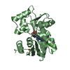
 Chitinophaga pinensis DSM 2588 (bacteria)
 X-RAY DIFFRACTION /
  Authors
 United States, 1items
  Citation
 Structure visualization
 Downloads & links
 Other downloads
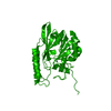


 PDBj
PDBj Assembly
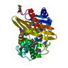
 Mass: 18.015 Da / Num. of mol.: 131 / Source method: isolated from a natural source / Formula: H2O
 Sample preparation
 Processing