 Movie
Movie Controller

[English] 日本語
 Yorodumi
- PDB-7k1s: The N-terminus of varicella-zoster virus glycoprotein B has a fun... -
+ Open data

- Basic information
| Entry | Database: PDB / ID: 7k1s | |||||||||||||||||||||||||||
|---|---|---|---|---|---|---|---|---|---|---|---|---|---|---|---|---|---|---|---|---|---|---|---|---|---|---|---|---|
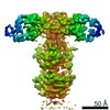
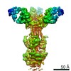
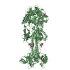
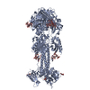
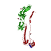
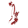
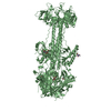
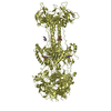
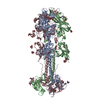
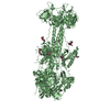
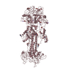
| Title | The N-terminus of varicella-zoster virus glycoprotein B has a functional role in fusion. | |||||||||||||||||||||||||||
 Components Components | Envelope glycoprotein B | |||||||||||||||||||||||||||
 Keywords Keywords | VIRAL PROTEIN / Herpesvirus / Varicella-Zoster Virus / Pathogenesis / Fusion / Glycoprotein B | |||||||||||||||||||||||||||
| Function / homology |  Function and homology information Function and homology informationviral envelope / symbiont entry into host cell / virion attachment to host cell Similarity search - Function | |||||||||||||||||||||||||||
| Biological species |  Human herpesvirus 3 (Varicella-zoster virus) Human herpesvirus 3 (Varicella-zoster virus) | |||||||||||||||||||||||||||
| Method | ELECTRON MICROSCOPY / single particle reconstruction / cryo EM / Resolution: 3.9 Å | |||||||||||||||||||||||||||
 Authors Authors | Oliver, S.L. | |||||||||||||||||||||||||||
| Funding support |  United States, 6items United States, 6items
| |||||||||||||||||||||||||||
 Citation Citation | Journal: PLoS Pathog / Year: 2021 Title: The N-terminus of varicella-zoster virus glycoprotein B has a functional role in fusion. Authors: Stefan L Oliver / Yi Xing / Dong-Hua Chen / Soung Hun Roh / Grigore D Pintilie / David A Bushnell / Marvin H Sommer / Edward Yang / Andrea Carfi / Wah Chiu / Ann M Arvin /  Abstract: Varicella-zoster virus (VZV) is a medically important alphaherpesvirus that induces fusion of the virion envelope and the cell membrane during entry, and between cells to form polykaryocytes within ...Varicella-zoster virus (VZV) is a medically important alphaherpesvirus that induces fusion of the virion envelope and the cell membrane during entry, and between cells to form polykaryocytes within infected tissues during pathogenesis. All members of the Herpesviridae, including VZV, have a conserved core fusion complex composed of glycoproteins, gB, gH and gL. The ectodomain of the primary fusogen, gB, has five domains, DI-V, of which DI contains the fusion loops needed for fusion function. We recently demonstrated that DIV is critical for fusion initiation, which was revealed by a 2.8Å structure of a VZV neutralizing mAb, 93k, bound to gB and mutagenesis of the gB-93k interface. To further assess the mechanism of mAb 93k neutralization, the binding site of a non-neutralizing mAb to gB, SG2, was compared to mAb 93k using single particle cryogenic electron microscopy (cryo-EM). The gB-SG2 interface partially overlapped with that of gB-93k but, unlike mAb 93k, mAb SG2 did not interact with the gB N-terminus, suggesting a potential role for the gB N-terminus in membrane fusion. The gB ectodomain structure in the absence of antibody was defined at near atomic resolution by single particle cryo-EM (3.9Å) of native, full-length gB purified from infected cells and by X-ray crystallography (2.4Å) of the transiently expressed ectodomain. Both structures revealed that the VZV gB N-terminus (aa72-114) was flexible based on the absence of visible structures in the cryo-EM or X-ray crystallography data but the presence of gB N-terminal peptides were confirmed by mass spectrometry. Notably, N-terminal residues 109KSQD112 were predicted to form a small α-helix and alanine substitution of these residues abolished cell-cell fusion in a virus-free assay. Importantly, transferring the 109AAAA112 mutation into the VZV genome significantly impaired viral propagation. These data establish a functional role for the gB N-terminus in membrane fusion broadly relevant to the Herpesviridae. | |||||||||||||||||||||||||||
| History |
|
- Structure visualization
| Movie |
Movie viewer |
|---|---|
| Structure viewer | Molecule: MolmilJmol/JSmol |
- Downloads & links
-Download
| PDBx/mmCIF format | 7k1s.cif.gz | 337.1 KB | Display | PDBx/mmCIF format |
|---|---|---|---|---|
| PDB format | pdb7k1s.ent.gz | 259.6 KB | Display | PDB format |
| PDBx/mmJSON format | 7k1s.json.gz | Tree view | PDBx/mmJSON format | |
| Others |  Other downloads Other downloads |
-Validation report
| Arichive directory | https://data.pdbj.org/pub/pdb/validation_reports/k1/7k1sftp://data.pdbj.org/pub/pdb/validation_reports/k1/7k1s | HTTPS FTP |
|---|
-Related structure data
| Related structure data |  22629MC M: map data used to model this data C: citing same article ( |
|---|---|
| Similar structure data |


-Links
 PDBj
PDBj- Assembly
| Deposited unit | 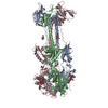
|
|---|---|
| 1 |
|
-Components
| #1: Protein | Mass: 105473.250 Da / Num. of mol.: 3 / Source method: isolated from a natural source Source: (natural) Human herpesvirus 3 (Varicella-zoster virus)References: UniProt: A0A076N502 #2: Polysaccharide | Source method: isolated from a genetically manipulated source #3: Sugar |   Type: D-saccharide, beta linking / Mass: 221.208 Da / Num. of mol.: 3 Type: D-saccharide, beta linking / Mass: 221.208 Da / Num. of mol.: 3Source method: isolated from a genetically manipulated source Formula: C8H15NO6 Has ligand of interest | N | Has protein modification | Y | |
|---|
-Experimental details
-Experiment
| Experiment | Method: ELECTRON MICROSCOPY |
|---|---|
| EM experiment | Aggregation state: PARTICLE / 3D reconstruction method: single particle reconstruction |
- Sample preparation
| Component | Name: Varicella-zoster virus glycoprotein B trimer. / Type: COMPLEX Details: The varicella-zoster virus glycoprotein B trimer was purified from infected MeWo cells. Entity ID: #1 / Source: NATURAL |
|---|---|
| Source (natural) | Organism: Human alphaherpesvirus 3 (Varicella-zoster virus) |
| Details of virus | Isolate: STRAIN |
| Buffer solution | pH: 7.4 |
| Specimen | Embedding applied: NO / Shadowing applied: NO / Staining applied: NO / Vitrification applied: YES |
| Vitrification | Cryogen name: ETHANE |
- Electron microscopy imaging
| Experimental equipment |  Model: Titan Krios / Image courtesy: FEI Company |
|---|---|
| Microscopy | Model: FEI TITAN KRIOS |
| Electron gun | Electron source:  FIELD EMISSION GUN / Accelerating voltage: 300 kV / Illumination mode: OTHER FIELD EMISSION GUN / Accelerating voltage: 300 kV / Illumination mode: OTHER |
| Electron lens | Mode: OTHER |
| Image recording | Electron dose: 7.5 e/Å2 / Film or detector model: GATAN K2 SUMMIT (4k x 4k) |
- Processing
| Software | Name: PHENIX / Version: 1.15.2_3472: / Classification: refinement | ||||||||||||||||||||||||
|---|---|---|---|---|---|---|---|---|---|---|---|---|---|---|---|---|---|---|---|---|---|---|---|---|---|
| EM software | Name: PHENIX / Category: model refinement | ||||||||||||||||||||||||
| CTF correction | Type: NONE | ||||||||||||||||||||||||
| 3D reconstruction | Resolution: 3.9 Å / Resolution method: FSC 0.143 CUT-OFF / Num. of particles: 349207 / Symmetry type: POINT | ||||||||||||||||||||||||
| Refine LS restraints |
|
