 Movie
Movie Controller


+ Open data

- Basic information
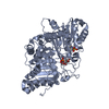
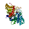
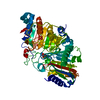
| Entry | Database: PDB / ID: 7jrb | ||||||
|---|---|---|---|---|---|---|---|
| Title | Phospholipase D engineered mutant | ||||||
 Components Components | Phospholipase D | ||||||
 Keywords Keywords | HYDROLASE / engineered mutant / pseudo-dimeric architecture / myo-inositol specificity | ||||||
| Function / homology |  Function and homology information Function and homology informationphosphatidyltransferase activity / cardiolipin biosynthetic process / phospholipase D / D-type glycerophospholipase activity / lipid catabolic process / extracellular region Similarity search - Function | ||||||
| Biological species |  Streptomyces antibioticus (bacteria) Streptomyces antibioticus (bacteria) | ||||||
| Method |  X-RAY DIFFRACTION / SYNCHROTRON / MOLECULAR REPLACEMENT / Resolution: 2.493 Å X-RAY DIFFRACTION / SYNCHROTRON / MOLECULAR REPLACEMENT / Resolution: 2.493 Å | ||||||
 Authors Authors | Vrielink, A. / Samantha, A. | ||||||
 Citation Citation | Journal: Biochem.J. / Year: 2021 Title: Structures of an engineered phospholipase D with specificity for secondary alcohol transphosphatidylation: insights into plasticity of substrate binding and activation. Authors: Samantha, A. / Damnjanovic, J. / Iwasaki, Y. / Nakano, H. / Vrielink, A. | ||||||
| History |
|
- Structure visualization
| Structure viewer | Molecule: MolmilJmol/JSmol |
|---|
- Downloads & links
-Download
| PDBx/mmCIF format | 7jrb.cif.gz | 108.5 KB | Display | PDBx/mmCIF format |
|---|---|---|---|---|
| PDB format | pdb7jrb.ent.gz | 80.2 KB | Display | PDB format |
| PDBx/mmJSON format | 7jrb.json.gz | Tree view | PDBx/mmJSON format | |
| Others |  Other downloads Other downloads |
-Validation report
| Arichive directory | https://data.pdbj.org/pub/pdb/validation_reports/jr/7jrbftp://data.pdbj.org/pub/pdb/validation_reports/jr/7jrb | HTTPS FTP |
|---|
-Related structure data
| Related structure data |  7jrcC  7jruC  7jrvC  7jrwC  7js5C  7js7C  2ze4S C: citing same article ( S: Starting model for refinement |
|---|---|
| Similar structure data |


-Links
 PDBj
PDBj- Assembly
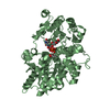
| Deposited unit | 
| ||||||||||||
|---|---|---|---|---|---|---|---|---|---|---|---|---|---|
| 1 |
| ||||||||||||
| Unit cell |
|
-Components
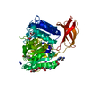
| #1: Protein | Mass: 54002.027 Da / Num. of mol.: 1 / Fragment: UNP residues 48-556 / Mutation: V107S,G186T,W187N,A211R,Y385R Source method: isolated from a genetically manipulated source Source: (gene. exp.) Streptomyces antibioticus (bacteria) / Production host: |
|---|---|
| #2: Chemical | ChemComp-MES /   Mass: 195.237 Da / Num. of mol.: 1 / Source method: obtained synthetically / Formula: C6H13NO4S / Comment: pH buffer*YM Mass: 195.237 Da / Num. of mol.: 1 / Source method: obtained synthetically / Formula: C6H13NO4S / Comment: pH buffer*YM |
| #3: Chemical | ChemComp-GOL /   Mass: 92.094 Da / Num. of mol.: 1 / Source method: obtained synthetically / Formula: C3H8O3 Mass: 92.094 Da / Num. of mol.: 1 / Source method: obtained synthetically / Formula: C3H8O3 |
| #4: Water | ChemComp-HOH /  Mass: 18.015 Da / Num. of mol.: 44 / Source method: isolated from a natural source / Formula: H2O Mass: 18.015 Da / Num. of mol.: 44 / Source method: isolated from a natural source / Formula: H2O |
| Has ligand of interest | N |
| Has protein modification | Y |
-Experimental details
-Experiment
| Experiment | Method: X-RAY DIFFRACTION / Number of used crystals: 1 |
|---|
- Sample preparation
| Crystal | Density Matthews: 2.27 Å3/Da / Density % sol: 45.81 % |
|---|---|
| Crystal grow | Temperature: 293 K / Method: vapor diffusion / pH: 6 / Details: 15% PEG6000, 100 mM MES, pH 6.0 |
-Data collection
| Diffraction | Mean temperature: 100 K / Serial crystal experiment: N |
|---|---|
| Diffraction source | Source: SYNCHROTRON / Site: Australian Synchrotron  / Beamline: MX2 / Wavelength: 0.953734 Å / Beamline: MX2 / Wavelength: 0.953734 Å |
| Detector | Type: DECTRIS EIGER X 16M / Detector: PIXEL / Date: Nov 18, 2017 |
| Radiation | Protocol: SINGLE WAVELENGTH / Monochromatic (M) / Laue (L): M / Scattering type: x-ray |
| Radiation wavelength | Wavelength: 0.953734 Å / Relative weight: 1 |
| Reflection | Resolution: 2.49→49.2 Å / Num. obs: 17644 / % possible obs: 99.4 % / Redundancy: 6.6 % / CC1/2: 0.998 / Rmerge(I) obs: 0.1 / Net I/σ(I): 13.2 |
| Reflection shell | Resolution: 2.49→2.58 Å / Rmerge(I) obs: 0.76 / Num. unique obs: 1641 / CC1/2: 0.849 |
- Processing
| Software |
| ||||||||||||||||||||||||
|---|---|---|---|---|---|---|---|---|---|---|---|---|---|---|---|---|---|---|---|---|---|---|---|---|---|
| Refinement | Method to determine structure: MOLECULAR REPLACEMENT Starting model: PDB entry 2ZE4 Resolution: 2.493→49.184 Å / Cross valid method: FREE R-VALUE Stereochemistry target values: GeoStd + Monomer Library + CDL v1.2
| ||||||||||||||||||||||||
| Displacement parameters | Biso mean: 50.63 Å2 | ||||||||||||||||||||||||
| Refinement step | Cycle: LAST / Resolution: 2.493→49.184 Å
| ||||||||||||||||||||||||
| Refine LS restraints |
| ||||||||||||||||||||||||
| LS refinement shell | Resolution: 2.49→2.58 Å
|
