Movie
Movie Controller

[English] 日本語
 Yorodumi
- PDB-7jm3: Full-length three-dimensional structure of the influenza A virus ... -
+ Open data

- Basic information
| Entry | Database: PDB / ID: 7jm3 | ||||||||||||
|---|---|---|---|---|---|---|---|---|---|---|---|---|---|
| Title | Full-length three-dimensional structure of the influenza A virus M1 protein and its organization into a matrix layer | ||||||||||||
 Components Components | Matrix protein 1 | ||||||||||||
 Keywords Keywords | VIRAL PROTEIN / influenza A / virus / matrix layer / M1 protein | ||||||||||||
| Function / homology |  Function and homology information Function and homology informationAssembly of Viral Components at the Budding Site / Influenza Infection / Fusion of the Influenza Virion to the Host Cell Endosome / Release / Budding / Packaging of Eight RNA Segments / Uncoating of the Influenza Virion / Entry of Influenza Virion into Host Cell via Endocytosis / Viral RNP Complexes in the Host Cell Nucleus / NEP/NS2 Interacts with the Cellular Export Machinery ...Assembly of Viral Components at the Budding Site / Influenza Infection / Fusion of the Influenza Virion to the Host Cell Endosome / Release / Budding / Packaging of Eight RNA Segments / Uncoating of the Influenza Virion / Entry of Influenza Virion into Host Cell via Endocytosis / Viral RNP Complexes in the Host Cell Nucleus / NEP/NS2 Interacts with the Cellular Export Machinery / Viral mRNA Translation / viral budding from plasma membrane / structural constituent of virion / host cell nucleus / virion membrane / RNA binding / extracellular region / plasma membrane Similarity search - Function | ||||||||||||
| Biological species |   Influenza A virus Influenza A virus | ||||||||||||
| Method | ELECTRON MICROSCOPY / helical reconstruction / cryo EM / Resolution: 3.4 Å | ||||||||||||
 Authors Authors | Su, Z. / Pintilie, G. / Selzer, L. / Chiu, W. / Kirkegaard, K. | ||||||||||||
| Funding support |  United States, 3items United States, 3items
| ||||||||||||
 Citation Citation | Journal: PLoS Biol / Year: 2020 Title: Full-length three-dimensional structure of the influenza A virus M1 protein and its organization into a matrix layer. Authors: Lisa Selzer / Zhaoming Su / Grigore D Pintilie / Wah Chiu / Karla Kirkegaard /  Abstract: Matrix proteins are encoded by many enveloped viruses, including influenza viruses, herpes viruses, and coronaviruses. Underneath the viral envelope of influenza virus, matrix protein 1 (M1) forms an ...Matrix proteins are encoded by many enveloped viruses, including influenza viruses, herpes viruses, and coronaviruses. Underneath the viral envelope of influenza virus, matrix protein 1 (M1) forms an oligomeric layer critical for particle stability and pH-dependent RNA genome release. However, high-resolution structures of full-length monomeric M1 and the matrix layer have not been available, impeding antiviral targeting and understanding of the pH-dependent transitions involved in cell entry. Here, purification and extensive mutagenesis revealed protein-protein interfaces required for the formation of multilayered helical M1 oligomers similar to those observed in virions exposed to the low pH of cell entry. However, single-layered helical oligomers with biochemical and ultrastructural similarity to those found in infectious virions before cell entry were observed upon mutation of a single amino acid. The highly ordered structure of the single-layered oligomers and their likeness to the matrix layer of intact virions prompted structural analysis by cryo-electron microscopy (cryo-EM). The resulting 3.4-Å-resolution structure revealed the molecular details of M1 folding and its organization within the single-shelled matrix. The solution of the full-length M1 structure, the identification of critical assembly interfaces, and the development of M1 assembly assays with purified proteins are crucial advances for antiviral targeting of influenza viruses. #1: Journal: Plos Biol. / Year: 2020Title: Full-length three-dimensional structure of the influenza A virus M1 protein and its organization into a matrix layer Authors: Selzer, L. / Su, Z. / Pintilie, G. / Chiu, W. / Kirkegaard, K. | ||||||||||||
| History |
|
- Structure visualization
| Movie |
Movie viewer |
|---|---|
| Structure viewer | Molecule: MolmilJmol/JSmol |
- Downloads & links
-Download
| PDBx/mmCIF format | 7jm3.cif.gz | 93.5 KB | Display | PDBx/mmCIF format |
|---|---|---|---|---|
| PDB format | pdb7jm3.ent.gz | 72.4 KB | Display | PDB format |
| PDBx/mmJSON format | 7jm3.json.gz | Tree view | PDBx/mmJSON format | |
| Others |  Other downloads Other downloads |
-Validation report
| Arichive directory | https://data.pdbj.org/pub/pdb/validation_reports/jm/7jm3ftp://data.pdbj.org/pub/pdb/validation_reports/jm/7jm3 | HTTPS FTP |
|---|
-Related structure data
| Related structure data |  22384MC M: map data used to model this data C: citing same article ( |
|---|---|
| Similar structure data |


-Links
 PDBj
PDBj


- Assembly
| Deposited unit | 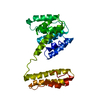
|
|---|---|
| 1 | x 60
|
| 2 |
|
| Symmetry | Helical symmetry: (Circular symmetry: 1 / Dyad axis: no / N subunits divisor: 1 / Num. of operations: 60 / Rise per n subunits: 1.96 Å / Rotation per n subunits: 17.1 °) |
-Components
| #1: Protein | Mass: 27827.152 Da / Num. of mol.: 1 / Mutation: V97K Source method: isolated from a genetically manipulated source Source: (gene. exp.) Influenza A virus (strain A/Puerto Rico/8/1934 H1N1)Strain: A/Puerto Rico/8/1934 H1N1 / Gene: M1, M / Production host:  |
|---|
-Experimental details
-Experiment
| Experiment | Method: ELECTRON MICROSCOPY |
|---|---|
| EM experiment | Aggregation state: FILAMENT / 3D reconstruction method: helical reconstruction |
- Sample preparation
| Component | Name: M1-V97K oligomer / Type: COMPLEX / Entity ID: all / Source: RECOMBINANT | |||||||||||||||
|---|---|---|---|---|---|---|---|---|---|---|---|---|---|---|---|---|
| Molecular weight | Value: 143 kDa/nm / Experimental value: NO | |||||||||||||||
| Source (natural) | Organism: Influenza A virus (A/Puerto Rico/8/1934(H1N1)) | |||||||||||||||
| Source (recombinant) | Organism: | |||||||||||||||
| Details of virus | Empty: NO / Enveloped: YES / Isolate: STRAIN / Type: VIRION | |||||||||||||||
| Buffer solution | pH: 7 | |||||||||||||||
| Buffer component |
| |||||||||||||||
| Specimen | Conc.: 5 mg/ml / Embedding applied: NO / Shadowing applied: NO / Staining applied: NO / Vitrification applied: YES Details: Influenza A virus matrix protein with V97K mutation assembled in vitro | |||||||||||||||
| Specimen support | Grid material: COPPER / Grid mesh size: 200 divisions/in. / Grid type: Quantifoil R1.2/1.3 | |||||||||||||||
| Vitrification | Instrument: FEI VITROBOT MARK IV / Cryogen name: ETHANE / Humidity: 100 % / Chamber temperature: 277 K / Details: Blot for 3 seconds once before plunging. |
- Electron microscopy imaging
| Experimental equipment |  Model: Titan Krios / Image courtesy: FEI Company |
|---|---|
| Microscopy | Model: FEI TITAN KRIOS |
| Electron gun | Electron source:  FIELD EMISSION GUN / Accelerating voltage: 300 kV / Illumination mode: FLOOD BEAM FIELD EMISSION GUN / Accelerating voltage: 300 kV / Illumination mode: FLOOD BEAM |
| Electron lens | Mode: BRIGHT FIELD / Nominal magnification: 165000 X / Calibrated defocus min: 300 nm / Calibrated defocus max: 4300 nm / Cs: 2.7 mm / C2 aperture diameter: 70 µm / Alignment procedure: BASIC |
| Specimen holder | Cryogen: NITROGEN / Specimen holder model: FEI TITAN KRIOS AUTOGRID HOLDER / Temperature (max): 78 K / Temperature (min): 78 K |
| Image recording | Average exposure time: 4 sec. / Electron dose: 36 e/Å2 / Detector mode: COUNTING / Film or detector model: GATAN K2 QUANTUM (4k x 4k) / Num. of grids imaged: 2 / Num. of real images: 440 |
| EM imaging optics | Energyfilter name: GIF Bioquantum / Energyfilter slit width: 20 eV |
| Image scans | Width: 3838 / Height: 3710 / Movie frames/image: 20 / Used frames/image: 1-20 |
- Processing
| EM software |
| ||||||||||||||||||||||||||||||||||||||||||||||||||
|---|---|---|---|---|---|---|---|---|---|---|---|---|---|---|---|---|---|---|---|---|---|---|---|---|---|---|---|---|---|---|---|---|---|---|---|---|---|---|---|---|---|---|---|---|---|---|---|---|---|---|---|
| CTF correction | Type: PHASE FLIPPING AND AMPLITUDE CORRECTION | ||||||||||||||||||||||||||||||||||||||||||||||||||
| Helical symmerty | Angular rotation/subunit: 17.1 ° / Axial rise/subunit: 1.96 Å / Axial symmetry: C1 | ||||||||||||||||||||||||||||||||||||||||||||||||||
| Particle selection | Num. of particles selected: 2268 | ||||||||||||||||||||||||||||||||||||||||||||||||||
| 3D reconstruction | Resolution: 3.4 Å / Resolution method: FSC 0.143 CUT-OFF / Num. of particles: 56602 / Algorithm: FOURIER SPACE / Num. of class averages: 200 / Symmetry type: HELICAL | ||||||||||||||||||||||||||||||||||||||||||||||||||
| Atomic model building | Protocol: AB INITIO MODEL / Space: REAL / Target criteria: Correlation coefficient | ||||||||||||||||||||||||||||||||||||||||||||||||||
| Atomic model building | PDB-ID: 1EA3 Accession code: 1EA3 / Source name: PDB / Type: experimental model |