 ムービー
ムービー コントローラー


+ データを開く

- 基本情報
基本情報
| 登録情報 | データベース: PDB / ID: 7jg1 | |||||||||||||||||||||||||||||||||||||||||||||||||||||||||
|---|---|---|---|---|---|---|---|---|---|---|---|---|---|---|---|---|---|---|---|---|---|---|---|---|---|---|---|---|---|---|---|---|---|---|---|---|---|---|---|---|---|---|---|---|---|---|---|---|---|---|---|---|---|---|---|---|---|---|
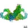
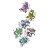
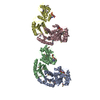
| タイトル | Dimeric Immunoglobin A (dIgA) | |||||||||||||||||||||||||||||||||||||||||||||||||||||||||
 要素 要素 |
| |||||||||||||||||||||||||||||||||||||||||||||||||||||||||
 キーワード キーワード | IMMUNE SYSTEM | |||||||||||||||||||||||||||||||||||||||||||||||||||||||||
| 機能・相同性 |  機能・相同性情報 機能・相同性情報Scavenging of heme from plasma / dimeric IgA immunoglobulin complex / secretory dimeric IgA immunoglobulin complex / pentameric IgM immunoglobulin complex / monomeric IgA immunoglobulin complex / secretory IgA immunoglobulin complex / IgA binding / Cell surface interactions at the vascular wall / glomerular filtration / peptidoglycan binding ...Scavenging of heme from plasma / dimeric IgA immunoglobulin complex / secretory dimeric IgA immunoglobulin complex / pentameric IgM immunoglobulin complex / monomeric IgA immunoglobulin complex / secretory IgA immunoglobulin complex / IgA binding / Cell surface interactions at the vascular wall / glomerular filtration / peptidoglycan binding / phosphatidylcholine binding / immunoglobulin receptor binding / immunoglobulin complex, circulating / positive regulation of respiratory burst / humoral immune response / complement activation, classical pathway / antigen binding / antibacterial humoral response / single-stranded DNA binding / protein-containing complex assembly / protein-macromolecule adaptor activity / adaptive immune response / innate immune response / protein homodimerization activity 類似検索 - 分子機能 | |||||||||||||||||||||||||||||||||||||||||||||||||||||||||
| 生物種 |  | |||||||||||||||||||||||||||||||||||||||||||||||||||||||||
| 手法 | 電子顕微鏡法 / 単粒子再構成法 / クライオ電子顕微鏡法 / 解像度: 3.3 Å | |||||||||||||||||||||||||||||||||||||||||||||||||||||||||
 データ登録者 データ登録者 | Kumar Bharathkar, S. / Parker, B.P. / Malyutin, A.G. / Stadtmueller, B.M. | |||||||||||||||||||||||||||||||||||||||||||||||||||||||||
| 資金援助 |  米国, 2件 米国, 2件
| |||||||||||||||||||||||||||||||||||||||||||||||||||||||||
 引用 引用 | ジャーナル: Elife / 年: 2020 タイトル: The structures of secretory and dimeric immunoglobulin A. 著者: Sonya Kumar Bharathkar / Benjamin W Parker / Andrey G Malyutin / Nandan Haloi / Kathryn E Huey-Tubman / Emad Tajkhorshid / Beth M Stadtmueller / 要旨: Secretory (S) Immunoglobulin (Ig) A is the predominant mucosal antibody, which binds pathogens and commensal microbes. SIgA is a polymeric antibody, typically containing two copies of IgA that ...Secretory (S) Immunoglobulin (Ig) A is the predominant mucosal antibody, which binds pathogens and commensal microbes. SIgA is a polymeric antibody, typically containing two copies of IgA that assemble with one joining-chain (JC) to form dimeric (d) IgA that is bound by the polymeric Ig-receptor ectodomain, called secretory component (SC). Here, we report the cryo-electron microscopy structures of murine SIgA and dIgA. Structures reveal two IgAs conjoined through four heavy-chain tailpieces and the JC that together form a β-sandwich-like fold. The two IgAs are bent and tilted with respect to each other, forming distinct concave and convex surfaces. In SIgA, SC is bound to one face, asymmetrically contacting both IgAs and JC. The bent and tilted arrangement of complex components limits the possible positions of both sets of antigen-binding fragments (Fabs) and preserves steric accessibility to receptor-binding sites, likely influencing antigen binding and effector functions. | |||||||||||||||||||||||||||||||||||||||||||||||||||||||||
| 履歴 |
|
- 構造の表示
構造の表示
| ムービー |
ムービービューア |
|---|---|
| 構造ビューア | 分子: MolmilJmol/JSmol |
- ダウンロードとリンク
-ダウンロード
| PDBx/mmCIF形式 | 7jg1.cif.gz | 344.4 KB | 表示 | PDBx/mmCIF形式 |
|---|---|---|---|---|
| PDB形式 | pdb7jg1.ent.gz | 279.9 KB | 表示 | PDB形式 |
| PDBx/mmJSON形式 | 7jg1.json.gz | ツリー表示 | PDBx/mmJSON形式 | |
| その他 |  その他のダウンロード その他のダウンロード |
-検証レポート
| 文書・要旨 | 7jg1_validation.pdf.gz | 1.6 MB | 表示 | wwPDB検証レポート |
|---|---|---|---|---|
| 文書・詳細版 | 7jg1_full_validation.pdf.gz | 1.6 MB | 表示 | |
| XML形式データ | 7jg1_validation.xml.gz | 46.9 KB | 表示 | |
| CIF形式データ | 7jg1_validation.cif.gz | 68.5 KB | 表示 | |
| アーカイブディレクトリ | https://data.pdbj.org/pub/pdb/validation_reports/jg/7jg1ftp://data.pdbj.org/pub/pdb/validation_reports/jg/7jg1 | HTTPS FTP |
-関連構造データ


-リンク
 PDBj
PDBj

- 集合体
集合体
| 登録構造単位 | 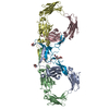
|
|---|---|
| 1 |
|
-要素
| #1: タンパク質 | 分子量: 37976.891 Da / 分子数: 4 / 由来タイプ: 組換発現 詳細: Genes encoding the mus musculus IgA HC constant region and the lambda LC constant region were fused with HC and LC variable region sequences to create complete HC and LC sequences. The HC and ...詳細: Genes encoding the mus musculus IgA HC constant region and the lambda LC constant region were fused with HC and LC variable region sequences to create complete HC and LC sequences. The HC and LC variable region is not modeled in this structure. Since authors have chosen not to provide complete sample sequence of the HC and LC variable region, the UNKs were not included in the sequence. 由来: (組換発現)  Homo sapiens (ヒト) / 参照: UniProt: Q99M22, UniProt: P01878*PLUS Homo sapiens (ヒト) / 参照: UniProt: Q99M22, UniProt: P01878*PLUS#2: タンパク質 | | 分子量: 15637.499 Da / 分子数: 1 / 由来タイプ: 組換発現 / 由来: (組換発現) Homo sapiens (ヒト) / 参照: UniProt: P01592#3: 多糖 | 2-acetamido-2-deoxy-beta-D-glucopyranose-(1-4)-2-acetamido-2-deoxy-beta-D-glucopyranose | #4: 糖 |   タイプ: D-saccharide, beta linking / 分子量: 221.208 Da / 分子数: 2 / 由来タイプ: 組換発現 / 式: C8H15NO6 タイプ: D-saccharide, beta linking / 分子量: 221.208 Da / 分子数: 2 / 由来タイプ: 組換発現 / 式: C8H15NO6研究の焦点であるリガンドがあるか | N | Has protein modification | Y | |
|---|
-実験情報
-実験
| 実験 | 手法: 電子顕微鏡法 |
|---|---|
| EM実験 | 試料の集合状態: PARTICLE / 3次元再構成法: 単粒子再構成法 |
- 試料調製
試料調製
| 構成要素 | 名称: Secretory Immunoglobin A / タイプ: COMPLEX / Entity ID: #1-#2 / 由来: RECOMBINANT |
|---|---|
| 由来(天然) | 生物種: |
| 由来(組換発現) | 生物種: Homo sapiens (ヒト) |
| 緩衝液 | pH: 7.4 |
| 試料 | 包埋: NO / シャドウイング: NO / 染色: NO / 凍結: YES |
| 急速凍結 | 装置: FEI VITROBOT MARK IV / 凍結剤: ETHANE / 湿度: 100 % / 凍結前の試料温度: 292 K 詳細: Wait time - 0s Drain time - 0s Blot time - 6s Blot Force - 5 |
- 電子顕微鏡撮影
| 実験機器 |  モデル: Titan Krios / 画像提供: FEI Company |
|---|---|
| 顕微鏡 | モデル: FEI TITAN KRIOS |
| 電子銃 | 電子線源:  FIELD EMISSION GUN / 加速電圧: 300 kV / 照射モード: FLOOD BEAM FIELD EMISSION GUN / 加速電圧: 300 kV / 照射モード: FLOOD BEAM |
| 電子レンズ | モード: BRIGHT FIELD / 倍率(公称値): 105000 X / 倍率(補正後): 59808 X / Cs: 2.7 mm / C2レンズ絞り径: 100 µm / アライメント法: COMA FREE |
| 試料ホルダ | 凍結剤: NITROGEN 試料ホルダーモデル: FEI TITAN KRIOS AUTOGRID HOLDER |
| 撮影 | 電子線照射量: 60 e/Å2 フィルム・検出器のモデル: GATAN K3 BIOQUANTUM (6k x 4k) |
| 電子光学装置 | エネルギーフィルター名称: GIF Bioquantum / エネルギーフィルタースリット幅: 20 eV |
| 画像スキャン | 横: 5760 / 縦: 4092 |
- 解析
解析
| ソフトウェア | 名称: PHENIX / バージョン: 1.17.1_3660: / 分類: 精密化 | ||||||||||||||||||||||||||||
|---|---|---|---|---|---|---|---|---|---|---|---|---|---|---|---|---|---|---|---|---|---|---|---|---|---|---|---|---|---|
| EMソフトウェア |
| ||||||||||||||||||||||||||||
| 画像処理 | 詳細: Super resolution images were collected. During motion correction, movies were binned by 2. | ||||||||||||||||||||||||||||
| CTF補正 | タイプ: PHASE FLIPPING AND AMPLITUDE CORRECTION | ||||||||||||||||||||||||||||
| 粒子像の選択 | 選択した粒子像数: 1808799 | ||||||||||||||||||||||||||||
| 3次元再構成 | 解像度: 3.3 Å / 解像度の算出法: FSC 0.143 CUT-OFF / 粒子像の数: 288823 / 対称性のタイプ: POINT | ||||||||||||||||||||||||||||
| 拘束条件 |
|
