 Movie
Movie Controller

[English] 日本語
 Yorodumi
- PDB-7ga9: PanDDA analysis group deposition -- Crystal Structure of MAP1LC3B... -
+ Open data

- Basic information
| Entry | Database: PDB / ID: 7ga9 | ||||||
|---|---|---|---|---|---|---|---|
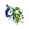
| Title | PanDDA analysis group deposition -- Crystal Structure of MAP1LC3B in complex with Z1198177230 | ||||||
 Components Components | Microtubule-associated proteins 1A/1B light chain 3B | ||||||
 Keywords Keywords | STRUCTURAL PROTEIN / SGC - Diamond I04-1 fragment screening / PanDDA / XChemExplorer | ||||||
| Function / homology |  Function and homology information Function and homology informationSARS-CoV-2 modulates autophagy / ceramide binding / phosphatidylethanolamine binding / Translation of Replicase and Assembly of the Replication Transcription Complex / TBC/RABGAPs / cellular response to nitrogen starvation / Receptor Mediated Mitophagy / Macroautophagy / organelle membrane / autophagosome membrane ...SARS-CoV-2 modulates autophagy / ceramide binding / phosphatidylethanolamine binding / Translation of Replicase and Assembly of the Replication Transcription Complex / TBC/RABGAPs / cellular response to nitrogen starvation / Receptor Mediated Mitophagy / Macroautophagy / organelle membrane / autophagosome membrane / autophagosome maturation / axoneme / autophagosome assembly / mitophagy / endomembrane system / autophagosome / Pexophagy / cellular response to starvation / PINK1-PRKN Mediated Mitophagy / macroautophagy / autophagy / mitochondrial membrane / KEAP1-NFE2L2 pathway / cytoplasmic vesicle / Translation of Replicase and Assembly of the Replication Transcription Complex / microtubule binding / microtubule / ubiquitin protein ligase binding / mitochondrion / cytosol Similarity search - Function | ||||||
| Biological species |  Homo sapiens (human) Homo sapiens (human) | ||||||
| Method |  X-RAY DIFFRACTION / SYNCHROTRON / FOURIER SYNTHESIS / molecular replacement / Resolution: 2.17 Å X-RAY DIFFRACTION / SYNCHROTRON / FOURIER SYNTHESIS / molecular replacement / Resolution: 2.17 Å | ||||||
 Authors Authors | Kumar, A. / Marples, P.G. / Tomlinson, C.W.E. / Fearon, D. / von-Delft, F. / Knapp, S. / Structural Genomics Consortium (SGC) | ||||||
| Funding support | European Union, 1items
| ||||||
 Citation Citation | Journal: To Be Published Title: PanDDA analysis group deposition Authors: Kumar, A. / Knapp, S. / Structural Genomics Consortium (SGC) | ||||||
| History |
|
- Structure visualization
| Structure viewer | Molecule: MolmilJmol/JSmol |
|---|
- Downloads & links
-Download
| PDBx/mmCIF format | 7ga9.cif.gz | 42 KB | Display | PDBx/mmCIF format |
|---|---|---|---|---|
| PDB format | pdb7ga9.ent.gz | 29.1 KB | Display | PDB format |
| PDBx/mmJSON format | 7ga9.json.gz | Tree view | PDBx/mmJSON format | |
| Others |  Other downloads Other downloads |
-Validation report
| Arichive directory | https://data.pdbj.org/pub/pdb/validation_reports/ga/7ga9ftp://data.pdbj.org/pub/pdb/validation_reports/ga/7ga9 | HTTPS FTP |
|---|
-Group deposition
| ID | G_1002273 (21 entries) |
|---|---|
| Title | PanDDA analysis group deposition |
| Type | changed state |
| Description | MAP1LC3B screened against the DSi-poised Fragment Library by X-ray Crystallography at the XChem facility of Diamond Light Source beamline I04-1 |


-Related structure data
| Related structure data | 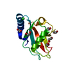 8q53S S: Starting model for refinement |
|---|---|
| Similar structure data |
-Links
 PDBj
PDBj


- Assembly
| Deposited unit | 
| ||||||||
|---|---|---|---|---|---|---|---|---|---|
| 1 |
| ||||||||
| Unit cell |
|
-Components
| #1: Protein | Mass: 14237.361 Da / Num. of mol.: 1 / Mutation: TruncationafterGly120 Source method: isolated from a genetically manipulated source Source: (gene. exp.) Homo sapiens (human) / Gene: MAP1LC3B, MAP1ALC3 / Production host:  | ||||||||
|---|---|---|---|---|---|---|---|---|---|
| #2: Chemical | ChemComp-K6U / ( Mass: 176.172 Da / Num. of mol.: 1 / Source method: obtained synthetically / Formula: C9H8N2O2 / Feature type: SUBJECT OF INVESTIGATION | ||||||||
| #3: Chemical | ChemComp-EDO /   Mass: 62.068 Da / Num. of mol.: 9 / Source method: obtained synthetically / Formula: C2H6O2 Mass: 62.068 Da / Num. of mol.: 9 / Source method: obtained synthetically / Formula: C2H6O2#4: Chemical | ChemComp-CL / |   Mass: 35.453 Da / Num. of mol.: 1 / Source method: obtained synthetically / Formula: Cl Mass: 35.453 Da / Num. of mol.: 1 / Source method: obtained synthetically / Formula: Cl#5: Water | ChemComp-HOH / |  Mass: 18.015 Da / Num. of mol.: 92 / Source method: isolated from a natural source / Formula: H2O Mass: 18.015 Da / Num. of mol.: 92 / Source method: isolated from a natural source / Formula: H2OHas ligand of interest | Y | Has protein modification | N | |
-Experimental details
-Experiment
| Experiment | Method: X-RAY DIFFRACTION / Number of used crystals: 1 |
|---|
- Sample preparation
| Crystal | Density Matthews: 2.22 Å3/Da / Density % sol: 44.57 % / Mosaicity: 0 ° |
|---|---|
| Crystal grow | Temperature: 293 K / Method: vapor diffusion, sitting drop / pH: 4.7 / Details: 36% PEG 8000, 0.1M acetate pH 4.7 |
-Data collection
| Diffraction | Mean temperature: 100 K | ||||||||||||||||||||||||||||||
|---|---|---|---|---|---|---|---|---|---|---|---|---|---|---|---|---|---|---|---|---|---|---|---|---|---|---|---|---|---|---|---|
| Diffraction source | Source: SYNCHROTRON / Site: Diamond  / Beamline: I04-1 / Wavelength: 0.92124 Å / Beamline: I04-1 / Wavelength: 0.92124 Å | ||||||||||||||||||||||||||||||
| Detector | Type: DECTRIS PILATUS 6M / Detector: PIXEL / Date: Feb 8, 2023 | ||||||||||||||||||||||||||||||
| Radiation | Protocol: SINGLE WAVELENGTH / Scattering type: x-ray | ||||||||||||||||||||||||||||||
| Radiation wavelength | Wavelength: 0.92124 Å / Relative weight: 1 | ||||||||||||||||||||||||||||||
| Reflection | Resolution: 2.17→30.49 Å / Num. obs: 7021 / % possible obs: 99.7 % / Redundancy: 13.3 % / CC1/2: 0.906 / Rmerge(I) obs: 0.199 / Rpim(I) all: 0.058 / Rrim(I) all: 0.208 / Net I/σ(I): 8.8 / Num. measured all: 93140 / Scaling rejects: 57 | ||||||||||||||||||||||||||||||
| Reflection shell | Diffraction-ID: 1
|
-Phasing
| Phasing | Method: molecular replacement |
|---|
- Processing
| Software |
| |||||||||||||||||||||||||||||||||||||||||||||||||||||||||||||||||||||||||||
|---|---|---|---|---|---|---|---|---|---|---|---|---|---|---|---|---|---|---|---|---|---|---|---|---|---|---|---|---|---|---|---|---|---|---|---|---|---|---|---|---|---|---|---|---|---|---|---|---|---|---|---|---|---|---|---|---|---|---|---|---|---|---|---|---|---|---|---|---|---|---|---|---|---|---|---|---|
| Refinement | Method to determine structure: FOURIER SYNTHESIS Starting model: 8Q53 Resolution: 2.17→30.51 Å / Cor.coef. Fo:Fc: 0.968 / Cor.coef. Fo:Fc free: 0.938 / SU B: 9.866 / SU ML: 0.226 / Cross valid method: THROUGHOUT / σ(F): 0 / ESU R: 0.352 / ESU R Free: 0.236 / Stereochemistry target values: MAXIMUM LIKELIHOOD Details: HYDROGENS HAVE BEEN ADDED IN THE RIDING POSITIONS U VALUES : REFINED INDIVIDUALLY
| |||||||||||||||||||||||||||||||||||||||||||||||||||||||||||||||||||||||||||
| Solvent computation | Ion probe radii: 0.8 Å / Shrinkage radii: 0.8 Å / VDW probe radii: 1.2 Å / Solvent model: MASK | |||||||||||||||||||||||||||||||||||||||||||||||||||||||||||||||||||||||||||
| Displacement parameters | Biso max: 156.73 Å2 / Biso mean: 56.978 Å2 / Biso min: 34.71 Å2
| |||||||||||||||||||||||||||||||||||||||||||||||||||||||||||||||||||||||||||
| Refinement step | Cycle: final / Resolution: 2.17→30.51 Å
| |||||||||||||||||||||||||||||||||||||||||||||||||||||||||||||||||||||||||||
| Refine LS restraints |
| |||||||||||||||||||||||||||||||||||||||||||||||||||||||||||||||||||||||||||
| LS refinement shell | Resolution: 2.171→2.227 Å / Total num. of bins used: 20
|
