 Movie
Movie Controller

[English] 日本語
 Yorodumi
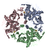
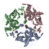
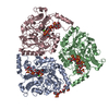
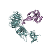
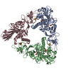
- PDB-7fhr: Crystal Structure of a Rieske Oxygenase from Cupriavidus metallidurans -
+ Open data

- Basic information
| Entry | Database: PDB / ID: 7fhr | ||||||
|---|---|---|---|---|---|---|---|
| Title | Crystal Structure of a Rieske Oxygenase from Cupriavidus metallidurans | ||||||
 Components Components | Putative Phthalate 4,5-dioxygenase, subunit alpha | ||||||
 Keywords Keywords | OXIDOREDUCTASE / dioxygenase / bacterial protein / metalloprotein / Rieske domain | ||||||
| Function / homology |  Function and homology information Function and homology informationphthalate 4,5-dioxygenase / phthalate 4,5-dioxygenase activity / catabolic process / 2 iron, 2 sulfur cluster binding / iron ion binding Similarity search - Function | ||||||
| Biological species |  Cupriavidus metallidurans CH34 (bacteria) Cupriavidus metallidurans CH34 (bacteria) | ||||||
| Method |  X-RAY DIFFRACTION / SYNCHROTRON / SAD / Resolution: 1.84 Å X-RAY DIFFRACTION / SYNCHROTRON / SAD / Resolution: 1.84 Å | ||||||
 Authors Authors | Mahto, J.K. / Dhankhar, P. / Kumar, P. | ||||||
| Funding support |  India, 1items India, 1items
| ||||||
 Citation Citation | Journal: J.Biol.Chem. / Year: 2021 Title: Molecular insights into substrate recognition and catalysis by phthalate dioxygenase from Comamonas testosteroni. Authors: Mahto, J.K. / Neetu, N. / Waghmode, B. / Kuatsjah, E. / Sharma, M. / Sircar, D. / Sharma, A.K. / Tomar, S. / Eltis, L.D. / Kumar, P. | ||||||
| History |
|
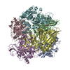
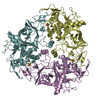
- Structure visualization
| Structure viewer | Molecule: MolmilJmol/JSmol |
|---|
- Downloads & links
-Download
| PDBx/mmCIF format | 7fhr.cif.gz | 196.2 KB | Display | PDBx/mmCIF format |
|---|---|---|---|---|
| PDB format | pdb7fhr.ent.gz | 153.9 KB | Display | PDB format |
| PDBx/mmJSON format | 7fhr.json.gz | Tree view | PDBx/mmJSON format | |
| Others |  Other downloads Other downloads |
-Validation report
| Arichive directory | https://data.pdbj.org/pub/pdb/validation_reports/fh/7fhrftp://data.pdbj.org/pub/pdb/validation_reports/fh/7fhr | HTTPS FTP |
|---|
-Related structure data


-Links
 PDBj
PDBj


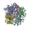
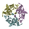
- Assembly
| Deposited unit | 
| ||||||||
|---|---|---|---|---|---|---|---|---|---|
| 1 | 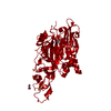
| ||||||||
| Unit cell |
|
-Components
-Protein , 1 types, 1 molecules A
| #1: Protein | Mass: 49756.129 Da / Num. of mol.: 1 Source method: isolated from a genetically manipulated source Source: (gene. exp.) Cupriavidus metallidurans CH34 (bacteria)Strain: CH34 / Gene: Rmet_5548 Production host: References: UniProt: Q1LBR9, phthalate 4,5-dioxygenase |
|---|
-Non-polymers , 7 types, 349 molecules 


| #2: Chemical | ChemComp-EDO /  Mass: 62.068 Da / Num. of mol.: 4 / Source method: obtained synthetically / Formula: C2H6O2 Mass: 62.068 Da / Num. of mol.: 4 / Source method: obtained synthetically / Formula: C2H6O2#3: Chemical | ChemComp-GLU / |  Type: L-peptide linking / Mass: 147.129 Da / Num. of mol.: 1 / Source method: obtained synthetically / Formula: C5H9NO4 Type: L-peptide linking / Mass: 147.129 Da / Num. of mol.: 1 / Source method: obtained synthetically / Formula: C5H9NO4#4: Chemical | ChemComp-GLY / |  Type: peptide linking / Mass: 75.067 Da / Num. of mol.: 1 / Source method: obtained synthetically / Formula: C2H5NO2 Type: peptide linking / Mass: 75.067 Da / Num. of mol.: 1 / Source method: obtained synthetically / Formula: C2H5NO2#5: Chemical | ChemComp-FES / |  Mass: 175.820 Da / Num. of mol.: 1 / Source method: obtained synthetically / Formula: Fe2S2 / Feature type: SUBJECT OF INVESTIGATION Mass: 175.820 Da / Num. of mol.: 1 / Source method: obtained synthetically / Formula: Fe2S2 / Feature type: SUBJECT OF INVESTIGATION#6: Chemical | ChemComp-FE2 / |  Mass: 55.845 Da / Num. of mol.: 1 / Source method: obtained synthetically / Formula: Fe / Feature type: SUBJECT OF INVESTIGATION Mass: 55.845 Da / Num. of mol.: 1 / Source method: obtained synthetically / Formula: Fe / Feature type: SUBJECT OF INVESTIGATION#7: Chemical | ChemComp-GOL / |  Mass: 92.094 Da / Num. of mol.: 1 / Source method: obtained synthetically / Formula: C3H8O3 Mass: 92.094 Da / Num. of mol.: 1 / Source method: obtained synthetically / Formula: C3H8O3#8: Water | ChemComp-HOH / | Mass: 18.015 Da / Num. of mol.: 340 / Source method: isolated from a natural source / Formula: H2O |
|---|
-Details
| Has ligand of interest | Y |
|---|
-Experimental details
-Experiment
| Experiment | Method: X-RAY DIFFRACTION / Number of used crystals: 1 |
|---|
- Sample preparation
| Crystal | Density Matthews: 2.41 Å3/Da / Density % sol: 48.99 % |
|---|---|
| Crystal grow | Temperature: 293 K / Method: vapor diffusion, sitting drop / Details: glycine, lysine, glutamate, PEG 4000 |
-Data collection
| Diffraction | Mean temperature: 100 K / Serial crystal experiment: N |
|---|---|
| Diffraction source | Source: SYNCHROTRON / Site: ESRF  / Beamline: ID30B / Wavelength: 1.7377 Å / Beamline: ID30B / Wavelength: 1.7377 Å |
| Detector | Type: DECTRIS PILATUS3 6M / Detector: PIXEL / Date: Nov 3, 2017 |
| Radiation | Monochromator: Fe / Protocol: SINGLE WAVELENGTH / Monochromatic (M) / Laue (L): M / Scattering type: x-ray |
| Radiation wavelength | Wavelength: 1.7377 Å / Relative weight: 1 |
| Reflection | Resolution: 1.84→77.59 Å / Num. obs: 40987 / % possible obs: 100 % / Redundancy: 2 % / CC1/2: 0.999 / Net I/σ(I): 16.3 |
| Reflection shell | Resolution: 1.84→1.88 Å / Num. unique obs: 2488 / CC1/2: 0.619 |
- Processing
| Software |
| |||||||||||||||||||||||||||||||||||||||||||||
|---|---|---|---|---|---|---|---|---|---|---|---|---|---|---|---|---|---|---|---|---|---|---|---|---|---|---|---|---|---|---|---|---|---|---|---|---|---|---|---|---|---|---|---|---|---|---|
| Refinement | Method to determine structure: SAD / Resolution: 1.84→58.71 Å / Cor.coef. Fo:Fc: 0.979 / Cor.coef. Fo:Fc free: 0.965 / SU B: 8.038 / SU ML: 0.116 / Cross valid method: THROUGHOUT / σ(F): 0 / ESU R: 0.125 / ESU R Free: 0.125 / Stereochemistry target values: MAXIMUM LIKELIHOOD / Details: U VALUES : WITH TLS ADDED
| |||||||||||||||||||||||||||||||||||||||||||||
| Solvent computation | Ion probe radii: 0.8 Å / Shrinkage radii: 0.8 Å / VDW probe radii: 1.2 Å / Solvent model: MASK | |||||||||||||||||||||||||||||||||||||||||||||
| Displacement parameters | Biso max: 141.58 Å2 / Biso mean: 43.974 Å2 / Biso min: 25.38 Å2
| |||||||||||||||||||||||||||||||||||||||||||||
| Refinement step | Cycle: final / Resolution: 1.84→58.71 Å
| |||||||||||||||||||||||||||||||||||||||||||||
| Refine LS restraints |
| |||||||||||||||||||||||||||||||||||||||||||||
| LS refinement shell | Resolution: 1.84→1.888 Å / Rfactor Rfree error: 0 / Total num. of bins used: 20
| |||||||||||||||||||||||||||||||||||||||||||||
| Refinement TLS params. | Method: refined / Origin x: -27.129 Å / Origin y: -17.396 Å / Origin z: -0.303 Å
| |||||||||||||||||||||||||||||||||||||||||||||
| Refinement TLS group |
|
