 Movie
Movie Controller


+ Open data

- Basic information
| Entry | Database: PDB / ID: 7ff1 | ||||||
|---|---|---|---|---|---|---|---|
| Title | Structure of C34E136G/N36 | ||||||
 Components Components |
| ||||||
 Keywords Keywords | VIRAL PROTEIN / HIV / envlope / 6HB | ||||||
| Function / homology | Retroviral envelope protein / Retroviral envelope protein GP41-like / viral envelope / host cell plasma membrane / virion membrane / structural molecule activity / plasma membrane / Env polyprotein Function and homology information Function and homology information | ||||||
| Biological species |   Human immunodeficiency virus 1 Human immunodeficiency virus 1 | ||||||
| Method |  X-RAY DIFFRACTION / SYNCHROTRON / MOLECULAR REPLACEMENT / Resolution: 1.689 Å X-RAY DIFFRACTION / SYNCHROTRON / MOLECULAR REPLACEMENT / Resolution: 1.689 Å | ||||||
 Authors Authors | Yu, D.W. / Qin, B. | ||||||
 Citation Citation | Journal: To Be Published Title: Structure of C34E136G/N36 Authors: Yu, D.W. / Qin, B. | ||||||
| History |
|
- Structure visualization
| Structure viewer | Molecule: MolmilJmol/JSmol |
|---|
- Downloads & links
-Download
| PDBx/mmCIF format | 7ff1.cif.gz | 102.5 KB | Display | PDBx/mmCIF format |
|---|---|---|---|---|
| PDB format | pdb7ff1.ent.gz | 79 KB | Display | PDB format |
| PDBx/mmJSON format | 7ff1.json.gz | Tree view | PDBx/mmJSON format | |
| Others |  Other downloads Other downloads |
-Validation report
| Summary document | 7ff1_validation.pdf.gz | 457.3 KB | Display | wwPDB validaton report |
|---|---|---|---|---|
| Full document | 7ff1_full_validation.pdf.gz | 464.3 KB | Display | |
| Data in XML | 7ff1_validation.xml.gz | 14.1 KB | Display | |
| Data in CIF | 7ff1_validation.cif.gz | 18.3 KB | Display | |
| Arichive directory | https://data.pdbj.org/pub/pdb/validation_reports/ff/7ff1ftp://data.pdbj.org/pub/pdb/validation_reports/ff/7ff1 | HTTPS FTP |
-Related structure data
| Related structure data | 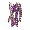 1aikS S: Starting model for refinement |
|---|---|
| Similar structure data |

-Links
 PDBj
PDBj


- Assembly
| Deposited unit | 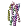
| ||||||||
|---|---|---|---|---|---|---|---|---|---|
| 1 |
| ||||||||
| Unit cell |
| ||||||||
| Components on special symmetry positions |
|
-Components
| #1: Protein/peptide | Mass: 4152.843 Da / Num. of mol.: 3 / Source method: obtained synthetically / Source: (synth.) Human immunodeficiency virus 1 / References: UniProt: C7F357#2: Protein/peptide | Mass: 4205.553 Da / Num. of mol.: 3 / Source method: obtained synthetically / Source: (synth.) Human immunodeficiency virus 1#3: Water | ChemComp-HOH / |  Mass: 18.015 Da / Num. of mol.: 133 / Source method: isolated from a natural source / Formula: H2O Mass: 18.015 Da / Num. of mol.: 133 / Source method: isolated from a natural source / Formula: H2OHas ligand of interest | N | Has protein modification | Y | |
|---|
-Experimental details
-Experiment
| Experiment | Method: X-RAY DIFFRACTION / Number of used crystals: 1 |
|---|
- Sample preparation
| Crystal | Density Matthews: 2.52 Å3/Da / Density % sol: 51.16 % |
|---|---|
| Crystal grow | Temperature: 294 K / Method: vapor diffusion, hanging drop / pH: 6.5 / Details: BIS-Tris,Polyethylene glycol monomethyl ether 5000 |
-Data collection
| Diffraction | Mean temperature: 100 K / Serial crystal experiment: N |
|---|---|
| Diffraction source | Source: SYNCHROTRON / Site: SSRF  / Beamline: BL19U1 / Wavelength: 0.987 Å / Beamline: BL19U1 / Wavelength: 0.987 Å |
| Detector | Type: DECTRIS PILATUS 6M / Detector: PIXEL / Date: Nov 19, 2017 |
| Radiation | Protocol: SINGLE WAVELENGTH / Monochromatic (M) / Laue (L): M / Scattering type: x-ray |
| Radiation wavelength | Wavelength: 0.987 Å / Relative weight: 1 |
| Reflection | Resolution: 1.689→43.74 Å / Num. obs: 28119 / % possible obs: 99.7 % / Redundancy: 6.628 % / CC1/2: 0.999 / Rmerge(I) obs: 0.043 / Net I/σ(I): 19.95 |
| Reflection shell | Resolution: 1.689→1.79 Å / Rmerge(I) obs: 0.976 / Mean I/σ(I) obs: 1.45 / Num. unique obs: 4478 / CC1/2: 0.757 / % possible all: 99.8 |
- Processing
| Software |
| ||||||||||||||||||||||||||||||||||||||||||||||||||||||||||||
|---|---|---|---|---|---|---|---|---|---|---|---|---|---|---|---|---|---|---|---|---|---|---|---|---|---|---|---|---|---|---|---|---|---|---|---|---|---|---|---|---|---|---|---|---|---|---|---|---|---|---|---|---|---|---|---|---|---|---|---|---|---|
| Refinement | Method to determine structure: MOLECULAR REPLACEMENT Starting model: 1AIK Resolution: 1.689→43.74 Å / SU ML: 0.26 / Cross valid method: THROUGHOUT / σ(F): 1.36 / Phase error: 29.51 / Stereochemistry target values: ML
| ||||||||||||||||||||||||||||||||||||||||||||||||||||||||||||
| Solvent computation | Shrinkage radii: 0.9 Å / VDW probe radii: 1.11 Å / Solvent model: FLAT BULK SOLVENT MODEL | ||||||||||||||||||||||||||||||||||||||||||||||||||||||||||||
| Displacement parameters | Biso max: 93.85 Å2 / Biso mean: 44.4355 Å2 / Biso min: 24.97 Å2 | ||||||||||||||||||||||||||||||||||||||||||||||||||||||||||||
| Refinement step | Cycle: final / Resolution: 1.689→43.74 Å
| ||||||||||||||||||||||||||||||||||||||||||||||||||||||||||||
| Refine LS restraints |
| ||||||||||||||||||||||||||||||||||||||||||||||||||||||||||||
| LS refinement shell | Refine-ID: X-RAY DIFFRACTION / Rfactor Rfree error: 0
|
