 Movie
Movie Controller


+ Open data

- Basic information
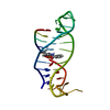
| Entry | Database: PDB / ID: 7feo | ||||||
|---|---|---|---|---|---|---|---|
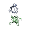
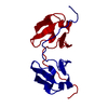
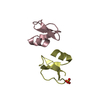
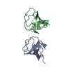
| Title | Crystal structure of AtMBD5 MBD domain | ||||||
 Components Components | Methyl-CpG-binding domain-containing protein 5 | ||||||
 Keywords Keywords | DNA BINDING PROTEIN / MBD domain / Arabidopsis thaliana / Structural Genomics / Structural Genomics Consortium / SGC | ||||||
| Function / homology |  Function and homology information Function and homology informationperinucleolar chromocenter / methyl-CpG binding / heterochromatin / enzyme binding / nucleus Similarity search - Function | ||||||
| Biological species |  | ||||||
| Method |  X-RAY DIFFRACTION / MOLECULAR REPLACEMENT / Resolution: 2.2 Å X-RAY DIFFRACTION / MOLECULAR REPLACEMENT / Resolution: 2.2 Å | ||||||
 Authors Authors | Zhou, M.Q. / Wu, Z.B. / Liu, K. / Min, J.R. / Structural Genomics Consortium (SGC) | ||||||
 Citation Citation | Journal: J.Mol.Biol. / Year: 2022 Title: Family-wide Characterization of Methylated DNA Binding Ability of Arabidopsis MBDs. Authors: Wu, Z. / Chen, S. / Zhou, M. / Jia, L. / Li, Z. / Zhang, X. / Min, J. / Liu, K. | ||||||
| History |
|
- Structure visualization
| Structure viewer | Molecule: MolmilJmol/JSmol |
|---|
- Downloads & links
-Download
| PDBx/mmCIF format | 7feo.cif.gz | 36 KB | Display | PDBx/mmCIF format |
|---|---|---|---|---|
| PDB format | pdb7feo.ent.gz | 23.4 KB | Display | PDB format |
| PDBx/mmJSON format | 7feo.json.gz | Tree view | PDBx/mmJSON format | |
| Others |  Other downloads Other downloads |
-Validation report
| Arichive directory | https://data.pdbj.org/pub/pdb/validation_reports/fe/7feoftp://data.pdbj.org/pub/pdb/validation_reports/fe/7feo | HTTPS FTP |
|---|
-Related structure data
| Related structure data |  7fefC  6c1tS S: Starting model for refinement C: citing same article ( |
|---|---|
| Similar structure data |


-Links
 PDBj
PDBj
- Assembly
| Deposited unit | 
| |||||||||
|---|---|---|---|---|---|---|---|---|---|---|
| 1 |
| |||||||||
| Unit cell |
| |||||||||
| Components on special symmetry positions |
|
-Components
| #1: Protein | Mass: 8230.116 Da / Num. of mol.: 2 Source method: isolated from a genetically manipulated source Source: (gene. exp.)  #2: Chemical |   Mass: 96.063 Da / Num. of mol.: 3 / Source method: obtained synthetically / Formula: SO4 Mass: 96.063 Da / Num. of mol.: 3 / Source method: obtained synthetically / Formula: SO4#3: Water | ChemComp-HOH / |  Mass: 18.015 Da / Num. of mol.: 12 / Source method: isolated from a natural source / Formula: H2O Mass: 18.015 Da / Num. of mol.: 12 / Source method: isolated from a natural source / Formula: H2OHas ligand of interest | N | |
|---|
-Experimental details
-Experiment
| Experiment | Method: X-RAY DIFFRACTION / Number of used crystals: 1 |
|---|
- Sample preparation
| Crystal | Density Matthews: 2.44 Å3/Da / Density % sol: 49.52 % |
|---|---|
| Crystal grow | Temperature: 291 K / Method: vapor diffusion, sitting drop / pH: 5.5 Details: 2M ammonium sulphate, 0.2M Sodium Chloride, 0.1M cacodylate pH5.5 |
-Data collection
| Diffraction | Mean temperature: 100 K / Serial crystal experiment: N |
|---|---|
| Diffraction source | Source: ROTATING ANODE / Type: RIGAKU FR-E SUPERBRIGHT / Wavelength: 1.54 Å |
| Detector | Type: RIGAKU SATURN A200 / Detector: CCD / Date: Mar 15, 2019 |
| Radiation | Protocol: SINGLE WAVELENGTH / Monochromatic (M) / Laue (L): M / Scattering type: x-ray |
| Radiation wavelength | Wavelength: 1.54 Å / Relative weight: 1 |
| Reflection | Resolution: 2.2→36.45 Å / Num. obs: 8605 / % possible obs: 99.91 % / Redundancy: 14.3 % / CC1/2: 1 / Net I/σ(I): 29.76 |
| Reflection shell | Resolution: 2.2→2.28 Å / Num. unique obs: 854 / CC1/2: 0.93 |
- Processing
| Software |
| ||||||||||||||||||||||||||||||||||||||||||||||||||||||||||||
|---|---|---|---|---|---|---|---|---|---|---|---|---|---|---|---|---|---|---|---|---|---|---|---|---|---|---|---|---|---|---|---|---|---|---|---|---|---|---|---|---|---|---|---|---|---|---|---|---|---|---|---|---|---|---|---|---|---|---|---|---|---|
| Refinement | Method to determine structure: MOLECULAR REPLACEMENT Starting model: 6c1t Resolution: 2.2→36.45 Å / Cor.coef. Fo:Fc: 0.951 / Cor.coef. Fo:Fc free: 0.941 / SU B: 6.884 / SU ML: 0.17 / Cross valid method: THROUGHOUT / σ(F): 0 / ESU R: 0.237 / ESU R Free: 0.201 / Stereochemistry target values: MAXIMUM LIKELIHOOD Details: HYDROGENS HAVE BEEN ADDED IN THE RIDING POSITIONS U VALUES : REFINED INDIVIDUALLY
| ||||||||||||||||||||||||||||||||||||||||||||||||||||||||||||
| Solvent computation | Ion probe radii: 0.8 Å / Shrinkage radii: 0.8 Å / VDW probe radii: 1.2 Å / Solvent model: MASK | ||||||||||||||||||||||||||||||||||||||||||||||||||||||||||||
| Displacement parameters | Biso max: 138.73 Å2 / Biso mean: 68.658 Å2 / Biso min: 32.43 Å2
| ||||||||||||||||||||||||||||||||||||||||||||||||||||||||||||
| Refinement step | Cycle: final / Resolution: 2.2→36.45 Å
| ||||||||||||||||||||||||||||||||||||||||||||||||||||||||||||
| Refine LS restraints |
| ||||||||||||||||||||||||||||||||||||||||||||||||||||||||||||
| LS refinement shell | Resolution: 2.2→2.257 Å / Rfactor Rfree error: 0 / Total num. of bins used: 20
|
