Method to determine structure: SAD / Resolution: 1.54→48.54 Å / Cor.coef. Fo:Fc: 0.974 / Cor.coef. Fo:Fc free: 0.968 / SU B: 3.317 / SU ML: 0.059 / Cross valid method: FREE R-VALUE / ESU R: 0.07 / ESU R Free: 0.07 Details: Hydrogens have been added in their riding positions
Rfactor
Num. reflection
% reflection
Rfree
0.1918
16770
4.919 %
Rwork
0.1709
324171
-
all
0.172
-
-
obs
-
340941
99.292 %
Solvent computation
Ion probe radii: 0.8 Å / Shrinkage radii: 0.8 Å / VDW probe radii: 1.2 Å / Solvent model: BABINET MODEL PLUS MASK
Displacement parameters
Biso mean: 29.102 Å2
Baniso -1
Baniso -2
Baniso -3
1-
-0.698 Å2
0 Å2
0.791 Å2
2-
-
-1.879 Å2
-0 Å2
3-
-
-
1.188 Å2
Refinement step
Cycle: LAST / Resolution: 1.54→48.54 Å
Protein
Nucleic acid
Ligand
Solvent
Total
Num. atoms
15741
0
104
1452
17297
Refine LS restraints
Refine-ID
Type
Dev ideal
Dev ideal target
Number
X-RAY DIFFRACTION
r_bond_refined_d
0.009
0.013
16452
X-RAY DIFFRACTION
r_bond_other_d
0.001
0.017
15341
X-RAY DIFFRACTION
r_angle_refined_deg
1.501
1.643
22319
X-RAY DIFFRACTION
r_angle_other_deg
1.416
1.579
35400
X-RAY DIFFRACTION
r_dihedral_angle_1_deg
6.849
5
2032
X-RAY DIFFRACTION
r_dihedral_angle_2_deg
36.817
23.819
830
X-RAY DIFFRACTION
r_dihedral_angle_3_deg
13.173
15
2799
X-RAY DIFFRACTION
r_dihedral_angle_4_deg
13.15
15
63
X-RAY DIFFRACTION
r_chiral_restr
0.08
0.2
2141
X-RAY DIFFRACTION
r_gen_planes_refined
0.008
0.02
18853
X-RAY DIFFRACTION
r_gen_planes_other
0.001
0.02
3797
X-RAY DIFFRACTION
r_nbd_refined
0.208
0.2
3184
X-RAY DIFFRACTION
r_symmetry_nbd_other
0.173
0.2
14326
X-RAY DIFFRACTION
r_nbtor_refined
0.171
0.2
7846
X-RAY DIFFRACTION
r_symmetry_nbtor_other
0.077
0.2
7497
X-RAY DIFFRACTION
r_xyhbond_nbd_refined
0.113
0.2
1262
X-RAY DIFFRACTION
r_symmetry_xyhbond_nbd_other
0.003
0.2
1
X-RAY DIFFRACTION
r_symmetry_nbd_refined
0.247
0.2
15
X-RAY DIFFRACTION
r_nbd_other
0.149
0.2
90
X-RAY DIFFRACTION
r_symmetry_xyhbond_nbd_refined
0.133
0.2
59
X-RAY DIFFRACTION
r_mcbond_it
0.983
2.109
8044
X-RAY DIFFRACTION
r_mcbond_other
0.983
2.109
8043
X-RAY DIFFRACTION
r_mcangle_it
1.477
3.16
10098
X-RAY DIFFRACTION
r_mcangle_other
1.477
3.161
10099
X-RAY DIFFRACTION
r_scbond_it
1.54
2.301
8408
X-RAY DIFFRACTION
r_scbond_other
1.54
2.301
8409
X-RAY DIFFRACTION
r_scangle_it
2.398
3.368
12219
X-RAY DIFFRACTION
r_scangle_other
2.398
3.368
12220
X-RAY DIFFRACTION
r_lrange_it
4.164
25.434
18962
X-RAY DIFFRACTION
r_lrange_other
4.073
24.886
18618
LS refinement shell
Resolution (Å)
Rfactor Rfree
Num. reflection Rfree
Rfactor Rwork
Num. reflection Rwork
Refine-ID
% reflection obs (%)
1.54-1.58
0.274
1236
0.272
23944
X-RAY DIFFRACTION
99.2511
1.58-1.623
0.262
1171
0.252
23446
X-RAY DIFFRACTION
99.3663
1.623-1.67
0.268
1148
0.239
22758
X-RAY DIFFRACTION
99.4219
1.67-1.722
0.227
1175
0.217
22064
X-RAY DIFFRACTION
99.5118
1.722-1.778
0.223
1073
0.203
21393
X-RAY DIFFRACTION
99.4511
1.778-1.841
0.22
1103
0.198
20675
X-RAY DIFFRACTION
99.3839
1.841-1.91
0.227
995
0.189
19958
X-RAY DIFFRACTION
99.2657
1.91-1.988
0.215
986
0.182
19169
X-RAY DIFFRACTION
99.2271
1.988-2.076
0.205
930
0.179
18447
X-RAY DIFFRACTION
99.2878
2.076-2.178
0.211
908
0.179
17610
X-RAY DIFFRACTION
99.255
2.178-2.295
0.206
876
0.171
16711
X-RAY DIFFRACTION
99.1431
2.295-2.435
0.192
860
0.166
15780
X-RAY DIFFRACTION
99.1657
2.435-2.603
0.206
797
0.163
14800
X-RAY DIFFRACTION
98.8528
2.603-2.811
0.18
701
0.158
13854
X-RAY DIFFRACTION
98.9665
2.811-3.079
0.189
616
0.158
12806
X-RAY DIFFRACTION
99.2825
3.079-3.442
0.175
588
0.164
11560
X-RAY DIFFRACTION
99.4027
3.442-3.974
0.169
560
0.154
10182
X-RAY DIFFRACTION
99.3985
3.974-4.865
0.154
463
0.141
8582
X-RAY DIFFRACTION
98.9498
4.865-6.872
0.165
359
0.153
6729
X-RAY DIFFRACTION
99.9295
6.872-48.54
0.16
225
0.154
3703
X-RAY DIFFRACTION
99.4934
Refinement TLS params.
Method: refined / Refine-ID: X-RAY DIFFRACTION
ID
L11 (°2)
L12 (°2)
L13 (°2)
L22 (°2)
L23 (°2)
L33 (°2)
S11 (Å °)
S12 (Å °)
S13 (Å °)
S21 (Å °)
S22 (Å °)
S23 (Å °)
S31 (Å °)
S32 (Å °)
S33 (Å °)
T11 (Å2)
T12 (Å2)
T13 (Å2)
T22 (Å2)
T23 (Å2)
T33 (Å2)
Origin x (Å)
Origin y (Å)
Origin z (Å)
1
0.1268
-0.0275
-0.0626
0.4401
0.2626
0.1953
-0.0079
0.0186
-0.032
-0.0206
-0.0325
0.1181
-0.0241
-0.0396
0.0404
0.0395
-0.006
0.026
0.0458
-0.0041
0.1196
-35.11
-11.858
0.71
2
0.3501
0.1363
0.061
0.3966
0.1287
0.1058
0.0175
-0.071
0.0735
0.0779
-0.0578
0.1277
-0.0039
-0.0036
0.0403
0.0919
0.0174
0.0735
0.0523
-0.0066
0.088
-30.553
12.264
24.898
3
0.0852
0.0166
0.0092
0.4094
-0.159
0.2282
0.0197
-0.0269
0.0165
0.0166
-0.0501
-0.0566
-0.0288
0.0331
0.0304
0.0716
-0.0132
0.0328
0.045
0.0012
0.0755
9.37
14.572
16.172
4
0.2353
0.5629
-0.2417
1.8547
-0.7931
0.4506
0.1196
-0.1331
-0.0952
0.312
-0.3131
-0.2345
-0.0883
0.1524
0.1936
0.1353
-0.0481
-0.0112
0.1022
0.0714
0.1109
12.915
-8.776
44.007
5
0.3996
0.0539
-0.0233
0.1349
-0.0402
0.0398
0.0184
-0.0042
-0.0609
0.0471
-0.0151
-0.0302
0.014
-0.0013
-0.0033
0.0933
0.011
0.0257
0.0171
0.0185
0.0908
6.523
-42.586
19.21
6
0.5628
-0.0641
0.1772
0.2128
-0.0773
0.1623
-0.0029
-0.1242
-0.0841
0.0432
0.0217
0.052
-0.0001
-0.0809
-0.0187
0.0936
-0.0212
0.0695
0.06
0.0124
0.0807
-27.291
-42.291
26.519
Refinement TLS group
ID
Refine-ID
Refine TLS-ID
Selection
Auth asym-ID
Auth seq-ID
1
X-RAY DIFFRACTION
1
ALL
A
23 - 360
2
X-RAY DIFFRACTION
2
ALL
A
361 - 681
3
X-RAY DIFFRACTION
3
ALL
B
23 - 284
4
X-RAY DIFFRACTION
4
ALL
B
285 - 681
5
X-RAY DIFFRACTION
5
ALL
C
23 - 353
6
X-RAY DIFFRACTION
6
ALL
C
354 - 681
+
About Yorodumi
-
News
-
Feb 9, 2022. New format data for meta-information of EMDB entries
New format data for meta-information of EMDB entries
Version 3 of the EMDB header file is now the official format.
The previous official version 1.9 will be removed from the archive.
In the structure databanks used in Yorodumi, some data are registered as the other names, "COVID-19 virus" and "2019-nCoV". Here are the details of the virus and the list of structure data.
Jan 31, 2019. EMDB accession codes are about to change! (news from PDBe EMDB page)
EMDB accession codes are about to change! (news from PDBe EMDB page)
The allocation of 4 digits for EMDB accession codes will soon come to an end. Whilst these codes will remain in use, new EMDB accession codes will include an additional digit and will expand incrementally as the available range of codes is exhausted. The current 4-digit format prefixed with “EMD-” (i.e. EMD-XXXX) will advance to a 5-digit format (i.e. EMD-XXXXX), and so on. It is currently estimated that the 4-digit codes will be depleted around Spring 2019, at which point the 5-digit format will come into force.
The EM Navigator/Yorodumi systems omit the EMD- prefix.
Related info.:Q: What is EMD? / ID/Accession-code notation in Yorodumi/EM Navigator
Yorodumi is a browser for structure data from EMDB, PDB, SASBDB, etc.
This page is also the successor to EM Navigator detail page, and also detail information page/front-end page for Omokage search.
The word "yorodu" (or yorozu) is an old Japanese word meaning "ten thousand". "mi" (miru) is to see.
Related info.:EMDB / PDB / SASBDB / Comparison of 3 databanks / Yorodumi Search / Aug 31, 2016. New EM Navigator & Yorodumi / Yorodumi Papers / Jmol/JSmol / Function and homology information / Changes in new EM Navigator and Yorodumi
 Movie
Movie Controller

 Yorodumi
 Open data
 Basic information
 Components
 Keywords
 Function and homology information
 Flavobacterium johnsoniae (bacteria)
 X-RAY DIFFRACTION /
  Authors
 Japan, 1items
  Citation
 Structure visualization
 Downloads & links
 Other downloads


 PDBj
PDBj


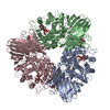
 Assembly


 Mass: 62.068 Da / Num. of mol.: 26 / Source method: obtained synthetically / Formula: C2H6O2
 Mass: 18.015 Da / Num. of mol.: 1452 / Source method: isolated from a natural source / Formula: H2O
 Sample preparation
 Processing