 Movie
Movie Controller

[English] 日本語
 Yorodumi
- PDB-7fdf: The E145S mutant of the regulatory domain of YeiE, a sulfite sens... -
+ Open data

- Basic information
| Entry | Database: PDB / ID: 7fdf | ||||||
|---|---|---|---|---|---|---|---|
| Title | The E145S mutant of the regulatory domain of YeiE, a sulfite sensing LysR-type transcriptional regulator from Cronobacter sakazakii (sulfate-bound form) | ||||||
 Components Components | LysR family transcriptional regulator | ||||||
 Keywords Keywords | DNA BINDING PROTEIN / LysR-type transcriptional regulator / LTTR / Cronobacter sakazakii / sulfite | ||||||
| Function / homology |  Function and homology information Function and homology informationtranscription cis-regulatory region binding / DNA-binding transcription factor activity / DNA-templated transcription Similarity search - Function | ||||||
| Biological species |  Cronobacter sakazakii (bacteria) Cronobacter sakazakii (bacteria) | ||||||
| Method |  X-RAY DIFFRACTION / SYNCHROTRON / MOLECULAR REPLACEMENT / Resolution: 2.05 Å X-RAY DIFFRACTION / SYNCHROTRON / MOLECULAR REPLACEMENT / Resolution: 2.05 Å | ||||||
 Authors Authors | Hong, S. / Ha, N.-C. | ||||||
| Funding support |  Korea, Republic Of, 1items Korea, Republic Of, 1items
| ||||||
 Citation Citation | Journal: Proc.Natl.Acad.Sci.USA / Year: 2022 Title: Crystal structures of YeiE from Cronobacter sakazakii and the role of sulfite tolerance in gram-negative bacteria. Authors: Hong, S. / Kim, J. / Cho, E. / Na, S. / Yoo, Y.J. / Cho, Y.H. / Ryu, S. / Ha, N.C. | ||||||
| History |
|
- Structure visualization
| Structure viewer | Molecule: MolmilJmol/JSmol |
|---|
- Downloads & links
-Download
| PDBx/mmCIF format | 7fdf.cif.gz | 66 KB | Display | PDBx/mmCIF format |
|---|---|---|---|---|
| PDB format | pdb7fdf.ent.gz | 37.6 KB | Display | PDB format |
| PDBx/mmJSON format | 7fdf.json.gz | Tree view | PDBx/mmJSON format | |
| Others |  Other downloads Other downloads |
-Validation report
| Arichive directory | https://data.pdbj.org/pub/pdb/validation_reports/fd/7fdfftp://data.pdbj.org/pub/pdb/validation_reports/fd/7fdf | HTTPS FTP |
|---|
-Related structure data
| Related structure data |  7erpSC 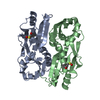 7erqC C: citing same article ( S: Starting model for refinement |
|---|---|
| Similar structure data |
-Links
 PDBj
PDBj- Assembly
| Deposited unit | 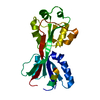
| ||||||||||||
|---|---|---|---|---|---|---|---|---|---|---|---|---|---|
| 1 | 
| ||||||||||||
| Unit cell |
|
-Components
| #1: Protein | Mass: 23221.934 Da / Num. of mol.: 1 / Mutation: E145S Source method: isolated from a genetically manipulated source Source: (gene. exp.) Cronobacter sakazakii (bacteria) / Gene: FZH93_18715, FZI36_01190, FZI41_05255, HZZ03_13430 / Production host: |
|---|---|
| #2: Chemical | ChemComp-SO4 /   Mass: 96.063 Da / Num. of mol.: 1 / Source method: obtained synthetically / Formula: SO4 / Feature type: SUBJECT OF INVESTIGATION Mass: 96.063 Da / Num. of mol.: 1 / Source method: obtained synthetically / Formula: SO4 / Feature type: SUBJECT OF INVESTIGATION |
| #3: Water | ChemComp-HOH /  Mass: 18.015 Da / Num. of mol.: 76 / Source method: isolated from a natural source / Formula: H2O Mass: 18.015 Da / Num. of mol.: 76 / Source method: isolated from a natural source / Formula: H2O |
| Has ligand of interest | Y |
-Experimental details
-Experiment
| Experiment | Method: X-RAY DIFFRACTION / Number of used crystals: 1 |
|---|
- Sample preparation
| Crystal | Density Matthews: 2.4 Å3/Da / Density % sol: 48.76 % |
|---|---|
| Crystal grow | Temperature: 287.15 K / Method: vapor diffusion, hanging drop / Details: 0.1M HEPES pH 7.5, 0.2M MgCl2, 19% (v/v) PEG 3350 |
-Data collection
| Diffraction | Mean temperature: 100.15 K / Serial crystal experiment: N |
|---|---|
| Diffraction source | Source: SYNCHROTRON / Site: PAL/PLS / Beamline: 5C (4A) / Wavelength: 1 Å |
| Detector | Type: DECTRIS EIGER X 9M / Detector: PIXEL / Date: Jul 13, 2021 |
| Radiation | Protocol: SINGLE WAVELENGTH / Monochromatic (M) / Laue (L): M / Scattering type: x-ray |
| Radiation wavelength | Wavelength: 1 Å / Relative weight: 1 |
| Reflection | Resolution: 2.05→50 Å / Num. obs: 14880 / % possible obs: 97.1 % / Redundancy: 22.2 % / Biso Wilson estimate: 20.77 Å2 / CC1/2: 1 / Rpim(I) all: 0.016 / Rrim(I) all: 0.083 / Net I/σ(I): 36.43 |
| Reflection shell | Resolution: 2.05→2.09 Å / Mean I/σ(I) obs: 5.625 / Num. unique obs: 685 / CC1/2: 0.955 / Rpim(I) all: 0.087 / Rrim(I) all: 0.429 / % possible all: 89.8 |
- Processing
| Software |
| ||||||||||||||||||||||||||||||||||||||||||||||||||||||||||||||||||||||||||||||||||||||||||||||||||||||||||||||||||||||||||||||
|---|---|---|---|---|---|---|---|---|---|---|---|---|---|---|---|---|---|---|---|---|---|---|---|---|---|---|---|---|---|---|---|---|---|---|---|---|---|---|---|---|---|---|---|---|---|---|---|---|---|---|---|---|---|---|---|---|---|---|---|---|---|---|---|---|---|---|---|---|---|---|---|---|---|---|---|---|---|---|---|---|---|---|---|---|---|---|---|---|---|---|---|---|---|---|---|---|---|---|---|---|---|---|---|---|---|---|---|---|---|---|---|---|---|---|---|---|---|---|---|---|---|---|---|---|---|---|---|
| Refinement | Method to determine structure: MOLECULAR REPLACEMENT Starting model: 7ERP Resolution: 2.05→35.59 Å / SU ML: 0.184 / Cross valid method: FREE R-VALUE / σ(F): 1.46 / Phase error: 19.6095 Stereochemistry target values: GeoStd + Monomer Library + CDL v1.2
| ||||||||||||||||||||||||||||||||||||||||||||||||||||||||||||||||||||||||||||||||||||||||||||||||||||||||||||||||||||||||||||||
| Solvent computation | Shrinkage radii: 0.9 Å / VDW probe radii: 1.11 Å / Solvent model: FLAT BULK SOLVENT MODEL | ||||||||||||||||||||||||||||||||||||||||||||||||||||||||||||||||||||||||||||||||||||||||||||||||||||||||||||||||||||||||||||||
| Displacement parameters | Biso mean: 23.33 Å2 | ||||||||||||||||||||||||||||||||||||||||||||||||||||||||||||||||||||||||||||||||||||||||||||||||||||||||||||||||||||||||||||||
| Refinement step | Cycle: LAST / Resolution: 2.05→35.59 Å
| ||||||||||||||||||||||||||||||||||||||||||||||||||||||||||||||||||||||||||||||||||||||||||||||||||||||||||||||||||||||||||||||
| Refine LS restraints |
| ||||||||||||||||||||||||||||||||||||||||||||||||||||||||||||||||||||||||||||||||||||||||||||||||||||||||||||||||||||||||||||||
| LS refinement shell |
|
