 Movie
Movie Controller


+ Open data

- Basic information
| Entry | Database: PDB / ID: 7fcj | ||||||
|---|---|---|---|---|---|---|---|
| Title | Zebrafish SIGIRR TIR domain mutant - C299S | ||||||
 Components Components | SIGIRR protein | ||||||
 Keywords Keywords | IMMUNE SYSTEM / receptor protein | ||||||
| Function / homology |  Function and homology information Function and homology informationnegative regulation of inflammatory response / cell surface / signal transduction / membrane Similarity search - Function | ||||||
| Biological species |  | ||||||
| Method |  X-RAY DIFFRACTION / SYNCHROTRON / MOLECULAR REPLACEMENT / Resolution: 1.88 Å X-RAY DIFFRACTION / SYNCHROTRON / MOLECULAR REPLACEMENT / Resolution: 1.88 Å | ||||||
 Authors Authors | Wang, X. / Zhou, J. | ||||||
 Citation Citation | Journal: Iscience / Year: 2022 Title: Structural basis of the IL-1 receptor TIR domain-mediated IL-1 signaling Authors: Zhou, J. / Xiao, Y. / Ren, Y. / Ge, J. / Wang, X. | ||||||
| History |
|
- Structure visualization
| Structure viewer | Molecule: MolmilJmol/JSmol |
|---|
- Downloads & links
-Download
| PDBx/mmCIF format | 7fcj.cif.gz | 47.8 KB | Display | PDBx/mmCIF format |
|---|---|---|---|---|
| PDB format | pdb7fcj.ent.gz | 31.1 KB | Display | PDB format |
| PDBx/mmJSON format | 7fcj.json.gz | Tree view | PDBx/mmJSON format | |
| Others |  Other downloads Other downloads |
-Validation report
| Arichive directory | https://data.pdbj.org/pub/pdb/validation_reports/fc/7fcjftp://data.pdbj.org/pub/pdb/validation_reports/fc/7fcj | HTTPS FTP |
|---|
-Related structure data
| Related structure data |  7fccSC 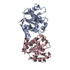 7fchC  7fclC 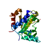 7fd3C S: Starting model for refinement C: citing same article ( |
|---|---|
| Similar structure data |
-Links
 PDBj
PDBj
- Assembly
| Deposited unit | 
| ||||||||
|---|---|---|---|---|---|---|---|---|---|
| 1 |
| ||||||||
| Unit cell |
|
-Components
| #1: Protein | Mass: 18403.182 Da / Num. of mol.: 1 / Mutation: C299S Source method: isolated from a genetically manipulated source Source: (gene. exp.)  |
|---|---|
| #2: Water | ChemComp-HOH /  Mass: 18.015 Da / Num. of mol.: 105 / Source method: isolated from a natural source / Formula: H2O Mass: 18.015 Da / Num. of mol.: 105 / Source method: isolated from a natural source / Formula: H2O |
-Experimental details
-Experiment
| Experiment | Method: X-RAY DIFFRACTION / Number of used crystals: 1 |
|---|
- Sample preparation
| Crystal | Density Matthews: 1.85 Å3/Da / Density % sol: 33.41 % Description: THE ENTRY CONTAINS FRIEDEL PAIRS IN I/F_PLUS/MINUS COLUMNS. |
|---|---|
| Crystal grow | Temperature: 291 K / Method: vapor diffusion, sitting drop / Details: 0.1 M CHES pH 9.5, 20% w/v PEG8000 |
-Data collection
| Diffraction | Mean temperature: 100 K / Serial crystal experiment: N |
|---|---|
| Diffraction source | Source: SYNCHROTRON / Site: SSRF  / Beamline: BL17U1 / Wavelength: 0.9796 Å / Beamline: BL17U1 / Wavelength: 0.9796 Å |
| Detector | Type: DECTRIS EIGER2 X 16M / Detector: PIXEL / Date: Oct 30, 2020 |
| Radiation | Protocol: SINGLE WAVELENGTH / Monochromatic (M) / Laue (L): M / Scattering type: x-ray |
| Radiation wavelength | Wavelength: 0.9796 Å / Relative weight: 1 |
| Reflection | Resolution: 1.88→50 Å / Num. obs: 10663 / % possible obs: 91.83 % / Redundancy: 9.2 % / Rmerge(I) obs: 0.13 / Net I/σ(I): 13 |
| Reflection shell | Resolution: 1.88→1.92 Å / Rmerge(I) obs: 0.537 / Num. unique obs: 631 |
- Processing
| Software |
| |||||||||||||||||||||||||||||||||||
|---|---|---|---|---|---|---|---|---|---|---|---|---|---|---|---|---|---|---|---|---|---|---|---|---|---|---|---|---|---|---|---|---|---|---|---|---|
| Refinement | Method to determine structure: MOLECULAR REPLACEMENT Starting model: 7FCC Resolution: 1.88→46.11 Å / SU ML: 0.23 / Cross valid method: THROUGHOUT / σ(F): 1.36 / Phase error: 26.12 / Stereochemistry target values: ML
| |||||||||||||||||||||||||||||||||||
| Solvent computation | Shrinkage radii: 0.9 Å / VDW probe radii: 1.11 Å / Solvent model: FLAT BULK SOLVENT MODEL | |||||||||||||||||||||||||||||||||||
| Displacement parameters | Biso max: 65.71 Å2 / Biso mean: 23.5894 Å2 / Biso min: 9.65 Å2 | |||||||||||||||||||||||||||||||||||
| Refinement step | Cycle: final / Resolution: 1.88→46.11 Å
| |||||||||||||||||||||||||||||||||||
| LS refinement shell | Refine-ID: X-RAY DIFFRACTION / Rfactor Rfree error: 0 / Total num. of bins used: 4
|
