Entry Database : PDB / ID : 7fcgTitle X-ray structure of high-strength hydrogel-grown FABP3 crystal soaked in 50% DMSO solution containing Indometacin Fatty acid-binding protein, heart Keywords / / / / Function / homology Function Domain/homology Component
/ / / / / / / / / / / / / / / / / / / / / / / / / / / / / / / / / / / / / / / / / / Biological species Homo sapiens (human)Method / / / Resolution : 1.19 Å Authors Sugiyama, S. / Kakinouchi, K. / Matsuoka, S. / Tsuchikawa, H. / Sonoyama, M. / Inoue, Y. / Hayashi, F. / Murata, M. Funding support Organization Grant number Country Japan Science and Technology JPMJTM19DC Japan Science and Technology JPMJER1005 Japan Society for the Promotion of Science (JSPS) 19K06588 Other private 09-003-005
Journal : To Be Published Title : X-ray structure of high-strength hydrogel-grown FABP3 crystal soaked in 50% DMSO solution containing IndometacinAuthors : Sugiyama, S. / Kakinouchi, K. / Matsuoka, S. / Tsuchikawa, H. / Sonoyama, M. / Inoue, Y. / Hayashi, F. / Murata, M. History Deposition Jul 14, 2021 Deposition site / Processing site Revision 1.0 Jul 20, 2022 Provider / Type Revision 1.1 Nov 29, 2023 Group / Refinement descriptionCategory / chem_comp_bond / pdbx_initial_refinement_model
Show all Show less
 Movie
Movie Controller

 Yorodumi
 Open data
 Basic information
 Components
 Keywords
 Function and homology information
 Homo sapiens (human)
 X-RAY DIFFRACTION /
  Authors
 Japan, 4items
  Citation
 Structure visualization
 Downloads & links
 Other downloads
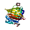
 PDBj
PDBj


 Assembly
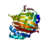

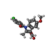
 Mass: 357.788 Da / Num. of mol.: 1 / Source method: obtained synthetically / Formula: C19H16ClNO4 / Feature type: SUBJECT OF INVESTIGATION / Comment: medication, antiinflammatory*YM
 Mass: 282.331 Da / Num. of mol.: 2 / Source method: obtained synthetically / Formula: C12H26O7 / Feature type: SUBJECT OF INVESTIGATION / Comment: precipitant*YM
 Mass: 18.015 Da / Num. of mol.: 181 / Source method: isolated from a natural source / Formula: H2O
 Sample preparation
 Processing