 Movie
Movie Controller


+ Open data

- Basic information
| Entry | Database: PDB / ID: 7fcb | ||||||
|---|---|---|---|---|---|---|---|
| Title | SptF 9 residues truncated mutant | ||||||
 Components Components | 2-oxoglutarate/Fe(II)-dependent dioxygenase SptF | ||||||
 Keywords Keywords | OXIDOREDUCTASE / SptF / alpha-ketoglutarate dependent oxygenase / biosynthesis | ||||||
| Function / homology |  Function and homology information Function and homology information | ||||||
| Biological species |  | ||||||
| Method |  X-RAY DIFFRACTION / SYNCHROTRON / MOLECULAR REPLACEMENT / Resolution: 1.4 Å X-RAY DIFFRACTION / SYNCHROTRON / MOLECULAR REPLACEMENT / Resolution: 1.4 Å | ||||||
 Authors Authors | Hui, T. / Mori, T. / Abe, I. | ||||||
 Citation Citation | Journal: Nat Commun / Year: 2022 Title: Molecular insights into the unusually promiscuous and catalytically versatile Fe(II)/ alpha-ketoglutarate-dependent oxygenase SptF. Authors: Tao, H. / Mori, T. / Chen, H. / Lyu, S. / Nonoyama, A. / Lee, S. / Abe, I. | ||||||
| History |
|
- Structure visualization
| Structure viewer | Molecule: MolmilJmol/JSmol |
|---|
- Downloads & links
-Download
| PDBx/mmCIF format | 7fcb.cif.gz | 92.2 KB | Display | PDBx/mmCIF format |
|---|---|---|---|---|
| PDB format | pdb7fcb.ent.gz | 54.3 KB | Display | PDB format |
| PDBx/mmJSON format | 7fcb.json.gz | Tree view | PDBx/mmJSON format | |
| Others |  Other downloads Other downloads |
-Validation report
| Arichive directory | https://data.pdbj.org/pub/pdb/validation_reports/fc/7fcbftp://data.pdbj.org/pub/pdb/validation_reports/fc/7fcb | HTTPS FTP |
|---|
-Related structure data
| Related structure data | 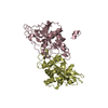 7eyrSC 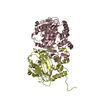 7eysC 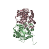 7eytC 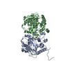 7eyuC 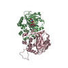 7eywC S: Starting model for refinement C: citing same article ( |
|---|---|
| Similar structure data |
-Links
 PDBj
PDBj


- Assembly
| Deposited unit | 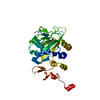
| ||||||||||||
|---|---|---|---|---|---|---|---|---|---|---|---|---|---|
| 1 | 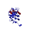
| ||||||||||||
| Unit cell |
| ||||||||||||
| Components on special symmetry positions |
|
-Components
| #1: Protein | Mass: 32727.922 Da / Num. of mol.: 1 Source method: isolated from a genetically manipulated source Source: (gene. exp.)  |
|---|---|
| #2: Chemical | ChemComp-OGA /   Mass: 147.086 Da / Num. of mol.: 1 / Source method: obtained synthetically / Formula: C4H5NO5 / Feature type: SUBJECT OF INVESTIGATION / Comment: inhibitor*YM Mass: 147.086 Da / Num. of mol.: 1 / Source method: obtained synthetically / Formula: C4H5NO5 / Feature type: SUBJECT OF INVESTIGATION / Comment: inhibitor*YM |
| #3: Chemical | ChemComp-FE2 /   Mass: 55.845 Da / Num. of mol.: 1 / Source method: obtained synthetically / Formula: Fe / Feature type: SUBJECT OF INVESTIGATION Mass: 55.845 Da / Num. of mol.: 1 / Source method: obtained synthetically / Formula: Fe / Feature type: SUBJECT OF INVESTIGATION |
| #4: Water | ChemComp-HOH /  Mass: 18.015 Da / Num. of mol.: 271 / Source method: isolated from a natural source / Formula: H2O Mass: 18.015 Da / Num. of mol.: 271 / Source method: isolated from a natural source / Formula: H2O |
| Has ligand of interest | Y |
-Experimental details
-Experiment
| Experiment | Method: X-RAY DIFFRACTION / Number of used crystals: 1 |
|---|
- Sample preparation
| Crystal | Density Matthews: 2.05 Å3/Da / Density % sol: 40.14 % |
|---|---|
| Crystal grow | Temperature: 293 K / Method: vapor diffusion, sitting drop / pH: 7 / Details: 0.2 M sodium formate (pH 7.0), 20% w/v PEG 3350 |
-Data collection
| Diffraction | Mean temperature: 100 K / Serial crystal experiment: N |
|---|---|
| Diffraction source | Source: SYNCHROTRON / Site: Photon Factory  / Beamline: BL-1A / Wavelength: 1.1 Å / Beamline: BL-1A / Wavelength: 1.1 Å |
| Detector | Type: DECTRIS EIGER X 4M / Detector: PIXEL / Date: Jun 27, 2021 |
| Radiation | Protocol: SINGLE WAVELENGTH / Monochromatic (M) / Laue (L): M / Scattering type: x-ray |
| Radiation wavelength | Wavelength: 1.1 Å / Relative weight: 1 |
| Reflection | Resolution: 1.4→45.1 Å / Num. obs: 52223 / % possible obs: 99.7 % / Redundancy: 3.8 % / Biso Wilson estimate: 12.45 Å2 / CC1/2: 0.999 / Rmerge(I) obs: 0.036 / Net I/σ(I): 12.4 |
| Reflection shell | Resolution: 1.4→1.42 Å / Redundancy: 3.9 % / Rmerge(I) obs: 0.374 / Mean I/σ(I) obs: 2.2 / Num. unique obs: 2577 / CC1/2: 0.854 / % possible all: 99.5 |
- Processing
| Software |
| |||||||||||||||||||||||||||||||||||||||||||||||||||||||||||||||||||||||||||||||||||||||||||||||||||||||||
|---|---|---|---|---|---|---|---|---|---|---|---|---|---|---|---|---|---|---|---|---|---|---|---|---|---|---|---|---|---|---|---|---|---|---|---|---|---|---|---|---|---|---|---|---|---|---|---|---|---|---|---|---|---|---|---|---|---|---|---|---|---|---|---|---|---|---|---|---|---|---|---|---|---|---|---|---|---|---|---|---|---|---|---|---|---|---|---|---|---|---|---|---|---|---|---|---|---|---|---|---|---|---|---|---|---|---|
| Refinement | Method to determine structure: MOLECULAR REPLACEMENT Starting model: 7EYR Resolution: 1.4→45.1 Å / SU ML: 0.1129 / Cross valid method: FREE R-VALUE / σ(F): 1.39 / Phase error: 17.6536 Stereochemistry target values: GeoStd + Monomer Library + CDL v1.2
| |||||||||||||||||||||||||||||||||||||||||||||||||||||||||||||||||||||||||||||||||||||||||||||||||||||||||
| Solvent computation | Shrinkage radii: 0.9 Å / VDW probe radii: 1.11 Å / Solvent model: FLAT BULK SOLVENT MODEL | |||||||||||||||||||||||||||||||||||||||||||||||||||||||||||||||||||||||||||||||||||||||||||||||||||||||||
| Displacement parameters | Biso mean: 17.1 Å2 | |||||||||||||||||||||||||||||||||||||||||||||||||||||||||||||||||||||||||||||||||||||||||||||||||||||||||
| Refinement step | Cycle: LAST / Resolution: 1.4→45.1 Å
| |||||||||||||||||||||||||||||||||||||||||||||||||||||||||||||||||||||||||||||||||||||||||||||||||||||||||
| Refine LS restraints |
| |||||||||||||||||||||||||||||||||||||||||||||||||||||||||||||||||||||||||||||||||||||||||||||||||||||||||
| LS refinement shell |
|
