 Movie
Movie Controller

[English] 日本語
 Yorodumi
- PDB-7fc9: Crystal structure of CmABCB1 in lipidic mesophase revealed by LCP-SFX -
+ Open data

- Basic information
| Entry | Database: PDB / ID: 7fc9 | ||||||
|---|---|---|---|---|---|---|---|
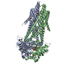
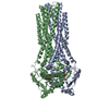
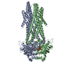
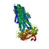
| Title | Crystal structure of CmABCB1 in lipidic mesophase revealed by LCP-SFX | ||||||
 Components Components | Probable ATP-dependent transporter ycf16 | ||||||
 Keywords Keywords | TRANSPORT PROTEIN / multi-drug exporter | ||||||
| Function / homology |  Function and homology information Function and homology informationoligopeptide export from mitochondrion / ABC-type oligopeptide transporter activity / mitochondrial inner membrane / ATP hydrolysis activity / ATP binding / metal ion binding Similarity search - Function | ||||||
| Biological species |  Cyanidioschyzon merolae (eukaryote) Cyanidioschyzon merolae (eukaryote) | ||||||
| Method |  X-RAY DIFFRACTION / FREE ELECTRON LASER / MOLECULAR REPLACEMENT / Resolution: 2.2 Å X-RAY DIFFRACTION / FREE ELECTRON LASER / MOLECULAR REPLACEMENT / Resolution: 2.2 Å | ||||||
 Authors Authors | Pan, D. / Oyama, R. / Sato, T. / Nakane, T. / Mizunuma, R. / Matsuoka, K. / Joti, Y. / Tono, K. / Nango, E. / Iwata, S. ...Pan, D. / Oyama, R. / Sato, T. / Nakane, T. / Mizunuma, R. / Matsuoka, K. / Joti, Y. / Tono, K. / Nango, E. / Iwata, S. / Nakatsu, T. / Kato, H. | ||||||
| Funding support |  Japan, 1items Japan, 1items
| ||||||
 Citation Citation | Journal: Iucrj / Year: 2022 Title: Crystal structure of CmABCB1 multi-drug exporter in lipidic mesophase revealed by LCP-SFX. Authors: Pan, D. / Oyama, R. / Sato, T. / Nakane, T. / Mizunuma, R. / Matsuoka, K. / Joti, Y. / Tono, K. / Nango, E. / Iwata, S. / Nakatsu, T. / Kato, H. | ||||||
| History |
|
- Structure visualization
| Structure viewer | Molecule: MolmilJmol/JSmol |
|---|
- Downloads & links
-Download
| PDBx/mmCIF format | 7fc9.cif.gz | 137 KB | Display | PDBx/mmCIF format |
|---|---|---|---|---|
| PDB format | pdb7fc9.ent.gz | 100.8 KB | Display | PDB format |
| PDBx/mmJSON format | 7fc9.json.gz | Tree view | PDBx/mmJSON format | |
| Others |  Other downloads Other downloads |
-Validation report
| Summary document | 7fc9_validation.pdf.gz | 1.6 MB | Display | wwPDB validaton report |
|---|---|---|---|---|
| Full document | 7fc9_full_validation.pdf.gz | 1.6 MB | Display | |
| Data in XML | 7fc9_validation.xml.gz | 12.7 KB | Display | |
| Data in CIF | 7fc9_validation.cif.gz | 19.8 KB | Display | |
| Arichive directory | https://data.pdbj.org/pub/pdb/validation_reports/fc/7fc9ftp://data.pdbj.org/pub/pdb/validation_reports/fc/7fc9 | HTTPS FTP |
-Related structure data
| Related structure data |  6a6mS S: Starting model for refinement |
|---|---|
| Similar structure data | |
| Experimental dataset #1 | Data reference: 10.11577/1835522 / Data set type: diffraction image data |


-Links
 PDBj
PDBj


- Assembly
| Deposited unit | 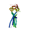
| ||||||||
|---|---|---|---|---|---|---|---|---|---|
| 1 | 
| ||||||||
| Unit cell |
|
-Components
-Protein , 1 types, 1 molecules A
| #1: Protein | Mass: 64175.621 Da / Num. of mol.: 1 / Mutation: Q147A, T381A Source method: isolated from a genetically manipulated source Source: (gene. exp.) Cyanidioschyzon merolae (strain 10D) (eukaryote)Strain: 10D / Gene: CYME_CMD148C / Production host:  Komagataella phaffii (fungus) / References: UniProt: M1VAN7 Komagataella phaffii (fungus) / References: UniProt: M1VAN7 |
|---|
-Non-polymers , 6 types, 163 molecules 


| #2: Chemical | ChemComp-ANP /  Mass: 506.196 Da / Num. of mol.: 1 / Source method: obtained synthetically / Formula: C10H17N6O12P3 / Feature type: SUBJECT OF INVESTIGATION / Comment: AMP-PNP, energy-carrying molecule analogue*YM Mass: 506.196 Da / Num. of mol.: 1 / Source method: obtained synthetically / Formula: C10H17N6O12P3 / Feature type: SUBJECT OF INVESTIGATION / Comment: AMP-PNP, energy-carrying molecule analogue*YM | ||||||||
|---|---|---|---|---|---|---|---|---|---|
| #3: Chemical | ChemComp-ZN /  Mass: 65.409 Da / Num. of mol.: 9 / Source method: obtained synthetically / Formula: Zn Mass: 65.409 Da / Num. of mol.: 9 / Source method: obtained synthetically / Formula: Zn#4: Chemical |  Mass: 24.305 Da / Num. of mol.: 2 / Source method: obtained synthetically / Formula: Mg / Feature type: SUBJECT OF INVESTIGATION Mass: 24.305 Da / Num. of mol.: 2 / Source method: obtained synthetically / Formula: Mg / Feature type: SUBJECT OF INVESTIGATION#5: Chemical |  Mass: 35.453 Da / Num. of mol.: 2 / Source method: obtained synthetically / Formula: Cl Mass: 35.453 Da / Num. of mol.: 2 / Source method: obtained synthetically / Formula: Cl#6: Chemical | ChemComp-ACT / |  Mass: 59.044 Da / Num. of mol.: 1 / Source method: obtained synthetically / Formula: C2H3O2 Mass: 59.044 Da / Num. of mol.: 1 / Source method: obtained synthetically / Formula: C2H3O2#7: Water | ChemComp-HOH / | Mass: 18.015 Da / Num. of mol.: 148 / Source method: isolated from a natural source / Formula: H2O |
-Details
| Has ligand of interest | Y |
|---|
-Experimental details
-Experiment
| Experiment | Method: X-RAY DIFFRACTION / Number of used crystals: 1 |
|---|
- Sample preparation
| Crystal | Density Matthews: 3.62 Å3/Da / Density % sol: 66.03 % |
|---|---|
| Crystal grow | Temperature: 293 K / Method: lipidic cubic phase Details: Micro-crystals were generated by incubation of 50 microL LCP containing the protein and 7.7 MAG/cholesterol in a 1.5 mL tube with 0.7 mL crystallization solution containing 26-30% 1,4- ...Details: Micro-crystals were generated by incubation of 50 microL LCP containing the protein and 7.7 MAG/cholesterol in a 1.5 mL tube with 0.7 mL crystallization solution containing 26-30% 1,4-butanediol, 0.1 M Tris pH 7.9, 0.2 M zinc acetate. |
-Data collection
| Diffraction | Mean temperature: 298 K / Serial crystal experiment: Y |
|---|---|
| Diffraction source | Source: FREE ELECTRON LASER / Site: SACLA / Beamline: BL3 / Wavelength: 1.771 Å |
| Detector | Type: MPCCD / Detector: CCD / Date: Nov 25, 2015 |
| Radiation | Protocol: SINGLE WAVELENGTH / Monochromatic (M) / Laue (L): M / Scattering type: x-ray |
| Radiation wavelength | Wavelength: 1.771 Å / Relative weight: 1 |
| Reflection | Resolution: 2.2→45.45 Å / Num. obs: 47845 / % possible obs: 100 % / Redundancy: 2149 % / CC star: 0.9996 / R split: 0.0567 / Net I/σ(I): 12.37 |
| Reflection shell | Resolution: 2.22→2.26 Å / Redundancy: 591 % / Mean I/σ(I) obs: 1.43 / Num. unique obs: 2335 / CC star: 0.7709 / R split: 0.8484 / % possible all: 100 |
| Serial crystallography sample delivery | Method: injection |
- Processing
| Software |
| |||||||||||||||||||||||||||||||||||||||||||||||||||||||||||||||||||||||||||||||||||||||||||||||||||||||||||||||||||||||||||||||||||||||||||||||||||||||||||||||||||||||||||||||||||||||||||||||||||||||||||||||||||||||||||||||||||||||
|---|---|---|---|---|---|---|---|---|---|---|---|---|---|---|---|---|---|---|---|---|---|---|---|---|---|---|---|---|---|---|---|---|---|---|---|---|---|---|---|---|---|---|---|---|---|---|---|---|---|---|---|---|---|---|---|---|---|---|---|---|---|---|---|---|---|---|---|---|---|---|---|---|---|---|---|---|---|---|---|---|---|---|---|---|---|---|---|---|---|---|---|---|---|---|---|---|---|---|---|---|---|---|---|---|---|---|---|---|---|---|---|---|---|---|---|---|---|---|---|---|---|---|---|---|---|---|---|---|---|---|---|---|---|---|---|---|---|---|---|---|---|---|---|---|---|---|---|---|---|---|---|---|---|---|---|---|---|---|---|---|---|---|---|---|---|---|---|---|---|---|---|---|---|---|---|---|---|---|---|---|---|---|---|---|---|---|---|---|---|---|---|---|---|---|---|---|---|---|---|---|---|---|---|---|---|---|---|---|---|---|---|---|---|---|---|---|---|---|---|---|---|---|---|---|---|---|---|---|---|---|---|---|
| Refinement | Method to determine structure: MOLECULAR REPLACEMENT Starting model: 6A6M Resolution: 2.2→20.001 Å / Cor.coef. Fo:Fc: 0.958 / Cor.coef. Fo:Fc free: 0.946 / WRfactor Rfree: 0.206 / WRfactor Rwork: 0.18 / Average fsc free: 0.9135 / Average fsc work: 0.921 / Cross valid method: FREE R-VALUE / ESU R: 0.18 / ESU R Free: 0.156 / Details: Hydrogens have not been used
| |||||||||||||||||||||||||||||||||||||||||||||||||||||||||||||||||||||||||||||||||||||||||||||||||||||||||||||||||||||||||||||||||||||||||||||||||||||||||||||||||||||||||||||||||||||||||||||||||||||||||||||||||||||||||||||||||||||||
| Solvent computation | Ion probe radii: 0.8 Å / Shrinkage radii: 0.8 Å / VDW probe radii: 1.2 Å / Solvent model: MASK BULK SOLVENT | |||||||||||||||||||||||||||||||||||||||||||||||||||||||||||||||||||||||||||||||||||||||||||||||||||||||||||||||||||||||||||||||||||||||||||||||||||||||||||||||||||||||||||||||||||||||||||||||||||||||||||||||||||||||||||||||||||||||
| Displacement parameters | Biso mean: 63.481 Å2
| |||||||||||||||||||||||||||||||||||||||||||||||||||||||||||||||||||||||||||||||||||||||||||||||||||||||||||||||||||||||||||||||||||||||||||||||||||||||||||||||||||||||||||||||||||||||||||||||||||||||||||||||||||||||||||||||||||||||
| Refinement step | Cycle: LAST / Resolution: 2.2→20.001 Å
| |||||||||||||||||||||||||||||||||||||||||||||||||||||||||||||||||||||||||||||||||||||||||||||||||||||||||||||||||||||||||||||||||||||||||||||||||||||||||||||||||||||||||||||||||||||||||||||||||||||||||||||||||||||||||||||||||||||||
| Refine LS restraints |
| |||||||||||||||||||||||||||||||||||||||||||||||||||||||||||||||||||||||||||||||||||||||||||||||||||||||||||||||||||||||||||||||||||||||||||||||||||||||||||||||||||||||||||||||||||||||||||||||||||||||||||||||||||||||||||||||||||||||
| LS refinement shell | Refine-ID: X-RAY DIFFRACTION / Total num. of bins used: 20
|
