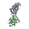
[English] 日本語
Yorodumi
- PDB-7f80: Co-crystal structure of Inhibitor compound MA-211 in complex with... -

+
Open data


ID or keywords:

Loading...

-
Basic information

Entry
Database: PDB / ID: 7f80
TitleCo-crystal structure of Inhibitor compound MA-211 in complex with human PPARdelta LBD
ComponentsPeroxisome proliferator-activated receptor delta
KeywordsPROTEIN BINDING/INHIBITOR / PROTEIN BINDING-INHIBITOR COMPLEX
Function / homology
Function and homology information


fat cell proliferation / positive regulation of fat cell proliferation / keratinocyte migration / linoleic acid binding / positive regulation of skeletal muscle tissue regeneration / axon ensheathment / regulation of skeletal muscle satellite cell proliferation / glucose transmembrane transport / positive regulation of myoblast proliferation / fatty acid catabolic process ...fat cell proliferation / positive regulation of fat cell proliferation / keratinocyte migration / linoleic acid binding / positive regulation of skeletal muscle tissue regeneration / axon ensheathment / regulation of skeletal muscle satellite cell proliferation / glucose transmembrane transport / positive regulation of myoblast proliferation / fatty acid catabolic process / Carnitine metabolism / negative regulation of myoblast differentiation / Regulation of pyruvate dehydrogenase (PDH) complex / Signaling by Retinoic Acid / nuclear steroid receptor activity / positive regulation of fatty acid metabolic process / cell-substrate adhesion / fatty acid beta-oxidation / negative regulation of cholesterol storage / cellular response to nutrient levels / decidualization / keratinocyte proliferation / positive regulation of fat cell differentiation / fatty acid transport / adipose tissue development / energy homeostasis / embryo implantation / hormone-mediated signaling pathway / cholesterol metabolic process / phosphatidylinositol 3-kinase/protein kinase B signal transduction / negative regulation of miRNA transcription / fatty acid metabolic process / generation of precursor metabolites and energy / apoptotic signaling pathway / wound healing / lipid metabolic process / transcription coactivator binding / DNA-binding transcription repressor activity, RNA polymerase II-specific / negative regulation of inflammatory response / Nuclear Receptor transcription pathway / glucose metabolic process / nuclear receptor activity / negative regulation of epithelial cell proliferation / sequence-specific double-stranded DNA binding / cellular response to hypoxia / DNA-binding transcription factor binding / cell population proliferation / positive regulation of phosphatidylinositol 3-kinase/protein kinase B signal transduction / cell differentiation / DNA-binding transcription factor activity, RNA polymerase II-specific / RNA polymerase II cis-regulatory region sequence-specific DNA binding / DNA-binding transcription factor activity / negative regulation of DNA-templated transcription / lipid binding / apoptotic process / positive regulation of gene expression / chromatin / regulation of transcription by RNA polymerase II / positive regulation of DNA-templated transcription / negative regulation of transcription by RNA polymerase II / positive regulation of transcription by RNA polymerase II / DNA binding / zinc ion binding / nucleoplasm / nucleus
Similarity search - Function
Peroxisome proliferator-activated receptor, beta / Peroxisome proliferator-activated receptor / Nuclear hormone receptor / Nuclear hormones receptors DNA-binding region signature. / Zinc finger, nuclear hormone receptor-type / Zinc finger, C4 type (two domains) / Nuclear hormone receptors DNA-binding domain profile. / c4 zinc finger in nuclear hormone receptors / Nuclear hormone receptor, ligand-binding domain / Nuclear hormone receptor-like domain superfamily ...Peroxisome proliferator-activated receptor, beta / Peroxisome proliferator-activated receptor / Nuclear hormone receptor / Nuclear hormones receptors DNA-binding region signature. / Zinc finger, nuclear hormone receptor-type / Zinc finger, C4 type (two domains) / Nuclear hormone receptors DNA-binding domain profile. / c4 zinc finger in nuclear hormone receptors / Nuclear hormone receptor, ligand-binding domain / Nuclear hormone receptor-like domain superfamily / Ligand-binding domain of nuclear hormone receptor / Nuclear receptor (NR) ligand-binding (LBD) domain profile. / Ligand binding domain of hormone receptors / Zinc finger, NHR/GATA-type
Similarity search - Domain/homology
Chem-1KF / Peroxisome proliferator-activated receptor delta
Similarity search - Component
Biological speciesHomo sapiens (human)
MethodX-RAY DIFFRACTION / SYNCHROTRON / MOLECULAR REPLACEMENT / Resolution: 2.8 Å
AuthorsLakshminarasimhan, A. / Rani, S.T. / Senaiar, R.S. / Krishnamurthy, N.
CitationJournal: To Be Published
Title: Co-crystal structure of Inhibitor compound in complex with human PPARdelta LBD
Authors: Lakshminarasimhan, A. / Rani, S.T. / Senaiar, R.S. / Krishnamurthy, N.
History
DepositionJun 30, 2021Deposition site: PDBJ / Processing site: PDBJ
Revision 1.0Jul 6, 2022Provider: repository / Type: Initial release
Revision 1.1Nov 29, 2023Group: Data collection / Refinement description
Category: chem_comp_atom / chem_comp_bond / pdbx_initial_refinement_model

-
Structure visualization

Structure viewerMolecule:
MolmilJmol/JSmol

Downloads & links

-
Assembly

Deposited unit
A: Peroxisome proliferator-activated receptor delta
B: Peroxisome proliferator-activated receptor delta
hetero molecules


Theoretical massNumber of molelcules
Total (without water)63,7824
Polymers62,8612
Non-polymers9212
Water00
1


  • Idetical with deposited unit
  • defined by author
  • Evidence: gel filtration
TypeNameSymmetry operationNumber
identity operation1_555x,y,z1
Buried area940 Å2
ΔGint-8 kcal/mol
Surface area24460 Å2
Unit cell
Length a, b, c (Å)39.687, 74.991, 96.341
Angle α, β, γ (deg.)90.00, 97.89, 90.00
Int Tables number4
Space group name H-MP1211

-
Components

#1: Protein Peroxisome proliferator-activated receptor delta / PPAR-delta / NUCI / Nuclear hormone receptor 1 / NUC1 / Nuclear receptor subfamily 1 group C member ...PPAR-delta / NUCI / Nuclear hormone receptor 1 / NUC1 / Nuclear receptor subfamily 1 group C member 2 / Peroxisome proliferator-activated receptor beta / PPAR-beta


Mass: 31430.535 Da / Num. of mol.: 2
Source method: isolated from a genetically manipulated source
Details: LIG / Source: (gene. exp.) Homo sapiens (human) / Gene: PPARD, NR1C2, PPARB / Production host: Escherichia phage EcSzw-2 (virus) / References: UniProt: Q03181
#2: Chemical ChemComp-1KF / (3R)-3-methyl-6-[2-[[5-methyl-2-[4-(trifluoromethyl)phenyl]imidazol-1-yl]methyl]phenoxy]hexanoic acid


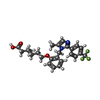
Mass: 460.489 Da / Num. of mol.: 2 / Source method: obtained synthetically / Formula: C25H27F3N2O3 / Feature type: SUBJECT OF INVESTIGATION
Has ligand of interestY

-
Experimental details

-
Experiment

ExperimentMethod: X-RAY DIFFRACTION / Number of used crystals: 1

-
Sample preparation

CrystalDensity Matthews: 2.43 Å3/Da / Density % sol: 49.41 %
Crystal growTemperature: 298 K / Method: vapor diffusion, hanging drop
Details: 40 mM Bis-Tris-propane (pH 8.0);14 % (w/v) polyethylene glycol (PEG) 8000;200 mM KCl, 6% propanol,1 mM CaCl2; 5 % n- Octyl-b-D- thioglucoside

-
Data collection

DiffractionMean temperature: 100 K / Serial crystal experiment: N
Diffraction sourceSource: SYNCHROTRON / Site: Australian Synchrotron / Beamline: MX2 / Wavelength: 1.1 Å
DetectorType: APEX II CCD / Detector: CCD / Date: Feb 15, 2016
RadiationProtocol: SINGLE WAVELENGTH / Monochromatic (M) / Laue (L): M / Scattering type: x-ray
Radiation wavelengthWavelength: 1.1 Å / Relative weight: 1
ReflectionResolution: 2.7→47.6 Å / Num. obs: 14162 / % possible obs: 90.3 % / Redundancy: 2.8 % / CC1/2: 0.88 / Rmerge(I) obs: 0.11 / Net I/σ(I): 6.9
Reflection shellResolution: 2.7→2.82 Å / Rmerge(I) obs: 0.562 / Num. unique obs: 1909 / CC1/2: 0.85

-
Processing

Software
NameVersionClassification
REFMAC5.7.0029refinement
CrystalCleardata reduction
Aimlessdata scaling
MOLREPphasing
RefinementMethod to determine structure: MOLECULAR REPLACEMENT
Starting model: 3TKM
Resolution: 2.8→47.6 Å / Cor.coef. Fo:Fc: 0.924 / Cor.coef. Fo:Fc free: 0.84 / SU B: 18.824 / SU ML: 0.381 / Cross valid method: THROUGHOUT / ESU R Free: 0.563 / Stereochemistry target values: MAXIMUM LIKELIHOOD / Details: HYDROGENS HAVE BEEN USED IF PRESENT IN THE INPUT
RfactorNum. reflection% reflectionSelection details
Rfree0.30039 586 5.1 %RANDOM
Rwork0.20427 ---
obs0.20915 10998 83.01 %-
Solvent computationIon probe radii: 0.8 Å / Shrinkage radii: 0.8 Å / VDW probe radii: 1.2 Å / Solvent model: MASK
Displacement parametersBiso mean: 37.224 Å2
Baniso -1Baniso -2Baniso -3
1--0.31 Å2-0 Å20.08 Å2
2--0.31 Å20 Å2
3----0 Å2
Refinement stepCycle: 1 / Resolution: 2.8→47.6 Å
ProteinNucleic acidLigandSolventTotal
Num. atoms4110 0 66 0 4176
Refine LS restraints
Refine-IDTypeDev idealDev ideal targetNumber
X-RAY DIFFRACTIONr_bond_refined_d0.0120.0194301
X-RAY DIFFRACTIONr_bond_other_d0.0030.0252
X-RAY DIFFRACTIONr_angle_refined_deg1.7141.9835812
X-RAY DIFFRACTIONr_angle_other_deg1.0153116
X-RAY DIFFRACTIONr_dihedral_angle_1_deg6.5285507
X-RAY DIFFRACTIONr_dihedral_angle_2_deg38.14624.136191
X-RAY DIFFRACTIONr_dihedral_angle_3_deg19.71215774
X-RAY DIFFRACTIONr_dihedral_angle_4_deg18.1591522
X-RAY DIFFRACTIONr_chiral_restr0.1020.2659
X-RAY DIFFRACTIONr_gen_planes_refined0.0070.0213160
X-RAY DIFFRACTIONr_gen_planes_other0.0020.0218
X-RAY DIFFRACTIONr_nbd_refined
X-RAY DIFFRACTIONr_nbd_other
X-RAY DIFFRACTIONr_nbtor_refined
X-RAY DIFFRACTIONr_nbtor_other
X-RAY DIFFRACTIONr_xyhbond_nbd_refined
X-RAY DIFFRACTIONr_xyhbond_nbd_other
X-RAY DIFFRACTIONr_metal_ion_refined
X-RAY DIFFRACTIONr_metal_ion_other
X-RAY DIFFRACTIONr_symmetry_vdw_refined
X-RAY DIFFRACTIONr_symmetry_vdw_other
X-RAY DIFFRACTIONr_symmetry_hbond_refined
X-RAY DIFFRACTIONr_symmetry_hbond_other
X-RAY DIFFRACTIONr_symmetry_metal_ion_refined
X-RAY DIFFRACTIONr_symmetry_metal_ion_other
X-RAY DIFFRACTIONr_mcbond_it
X-RAY DIFFRACTIONr_mcbond_other
X-RAY DIFFRACTIONr_mcangle_it
X-RAY DIFFRACTIONr_mcangle_other
X-RAY DIFFRACTIONr_scbond_it
X-RAY DIFFRACTIONr_scbond_other
X-RAY DIFFRACTIONr_scangle_it
X-RAY DIFFRACTIONr_scangle_other
X-RAY DIFFRACTIONr_long_range_B_refined
X-RAY DIFFRACTIONr_long_range_B_other
X-RAY DIFFRACTIONr_rigid_bond_restr
X-RAY DIFFRACTIONr_sphericity_free
X-RAY DIFFRACTIONr_sphericity_bonded
LS refinement shellResolution: 2.8→2.872 Å / Total num. of bins used: 20
RfactorNum. reflection% reflection
Rfree0.438 54 -
Rwork0.239 825 -
obs--84.28 %

+
About Yorodumi

-
News

-
Feb 9, 2022. New format data for meta-information of EMDB entries

New format data for meta-information of EMDB entries

  • Version 3 of the EMDB header file is now the official format.
  • The previous official version 1.9 will be removed from the archive.

Related info.:EMDB header

External links:wwPDB to switch to version 3 of the EMDB data model

-
Aug 12, 2020. Covid-19 info

Covid-19 info

URL: https://pdbjlvh1.pdbj.org/emnavi/covid19.php

New page: Covid-19 featured information page in EM Navigator.

Related info.:Covid-19 info / Mar 5, 2020. Novel coronavirus structure data

+
Mar 5, 2020. Novel coronavirus structure data

Novel coronavirus structure data

Related info.:Yorodumi Speices / Aug 12, 2020. Covid-19 info

External links:COVID-19 featured content - PDBj / Molecule of the Month (242):Coronavirus Proteases

+
Jan 31, 2019. EMDB accession codes are about to change! (news from PDBe EMDB page)

EMDB accession codes are about to change! (news from PDBe EMDB page)

  • The allocation of 4 digits for EMDB accession codes will soon come to an end. Whilst these codes will remain in use, new EMDB accession codes will include an additional digit and will expand incrementally as the available range of codes is exhausted. The current 4-digit format prefixed with “EMD-” (i.e. EMD-XXXX) will advance to a 5-digit format (i.e. EMD-XXXXX), and so on. It is currently estimated that the 4-digit codes will be depleted around Spring 2019, at which point the 5-digit format will come into force.
  • The EM Navigator/Yorodumi systems omit the EMD- prefix.

Related info.:Q: What is EMD? / ID/Accession-code notation in Yorodumi/EM Navigator

External links:EMDB Accession Codes are Changing Soon! / Contact to PDBj

+
Jul 12, 2017. Major update of PDB

Major update of PDB

  • wwPDB released updated PDB data conforming to the new PDBx/mmCIF dictionary.
  • This is a major update changing the version number from 4 to 5, and with Remediation, in which all the entries are updated.
  • In this update, many items about electron microscopy experimental information are reorganized (e.g. em_software).
  • Now, EM Navigator and Yorodumi are based on the updated data.

External links:wwPDB Remediation / Enriched Model Files Conforming to OneDep Data Standards Now Available in the PDB FTP Archive

-
Yorodumi

Thousand views of thousand structures

  • Yorodumi is a browser for structure data from EMDB, PDB, SASBDB, etc.
  • This page is also the successor to EM Navigator detail page, and also detail information page/front-end page for Omokage search.
  • The word "yorodu" (or yorozu) is an old Japanese word meaning "ten thousand". "mi" (miru) is to see.

Related info.:EMDB / PDB / SASBDB / Comparison of 3 databanks / Yorodumi Search / Aug 31, 2016. New EM Navigator & Yorodumi / Yorodumi Papers / Jmol/JSmol / Function and homology information / Changes in new EM Navigator and Yorodumi

Read more