 Movie
Movie Controller

[English] 日本語
 Yorodumi
- PDB-7f61: Crystal structure of human histamine receptor H3R in complex with... -
+ Open data

- Basic information
| Entry | Database: PDB / ID: 7f61 | |||||||||||||||||||||
|---|---|---|---|---|---|---|---|---|---|---|---|---|---|---|---|---|---|---|---|---|---|---|
| Title | Crystal structure of human histamine receptor H3R in complex with antagonist PF03654746 | |||||||||||||||||||||
 Components Components |
| |||||||||||||||||||||
 Keywords Keywords | MEMBRANE PROTEIN / GPCR / histamine receptor | |||||||||||||||||||||
| Function / homology |  Function and homology information Function and homology informationHistamine receptors / histamine receptor activity / neurotransmitter receptor activity / adenylate cyclase-inhibiting G protein-coupled acetylcholine receptor signaling pathway / neurotransmitter secretion / G protein-coupled receptor signaling pathway, coupled to cyclic nucleotide second messenger / electron transport chain / adenylate cyclase-activating G protein-coupled receptor signaling pathway / presynapse / chemical synaptic transmission ...Histamine receptors / histamine receptor activity / neurotransmitter receptor activity / adenylate cyclase-inhibiting G protein-coupled acetylcholine receptor signaling pathway / neurotransmitter secretion / G protein-coupled receptor signaling pathway, coupled to cyclic nucleotide second messenger / electron transport chain / adenylate cyclase-activating G protein-coupled receptor signaling pathway / presynapse / chemical synaptic transmission / periplasmic space / electron transfer activity / iron ion binding / heme binding / synapse / dendrite / plasma membrane Similarity search - Function | |||||||||||||||||||||
| Biological species |  Homo sapiens (human) Homo sapiens (human) | |||||||||||||||||||||
| Method |  X-RAY DIFFRACTION / SYNCHROTRON / MOLECULAR REPLACEMENT / Resolution: 2.6 Å X-RAY DIFFRACTION / SYNCHROTRON / MOLECULAR REPLACEMENT / Resolution: 2.6 Å | |||||||||||||||||||||
 Authors Authors | Peng, X. / Zhang, H. | |||||||||||||||||||||
| Funding support |  China, 6items China, 6items
| |||||||||||||||||||||
 Citation Citation | Journal: Nat Commun / Year: 2022 Title: Structural basis for recognition of antihistamine drug by human histamine receptor. Authors: Peng, X. / Yang, L. / Liu, Z. / Lou, S. / Mei, S. / Li, M. / Chen, Z. / Zhang, H. | |||||||||||||||||||||
| History |
|
- Structure visualization
| Structure viewer | Molecule: MolmilJmol/JSmol |
|---|
- Downloads & links
-Download
| PDBx/mmCIF format | 7f61.cif.gz | 101 KB | Display | PDBx/mmCIF format |
|---|---|---|---|---|
| PDB format | pdb7f61.ent.gz | 72.3 KB | Display | PDB format |
| PDBx/mmJSON format | 7f61.json.gz | Tree view | PDBx/mmJSON format | |
| Others |  Other downloads Other downloads |
-Validation report
| Arichive directory | https://data.pdbj.org/pub/pdb/validation_reports/f6/7f61ftp://data.pdbj.org/pub/pdb/validation_reports/f6/7f61 | HTTPS FTP |
|---|
-Related structure data
| Related structure data |  4u15S S: Starting model for refinement |
|---|---|
| Similar structure data |
-Links
 PDBj
PDBj


- Assembly
| Deposited unit | 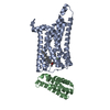
| ||||||||
|---|---|---|---|---|---|---|---|---|---|
| 1 |
| ||||||||
| Unit cell |
|
-Components
| #1: Protein | Mass: 45009.184 Da / Num. of mol.: 1 / Mutation: S121K Source method: isolated from a genetically manipulated source Source: (gene. exp.) Homo sapiens (human) / Gene: HRH3, GPCR97 / Production host:   Spodoptera frugiperda (fall armyworm) / References: UniProt: Q9Y5N1 Spodoptera frugiperda (fall armyworm) / References: UniProt: Q9Y5N1 |
|---|---|
| #2: Protein | Mass: 16831.879 Da / Num. of mol.: 1 / Mutation: M7W, H102I, R106L Source method: isolated from a genetically manipulated source Source: (gene. exp.) Spodoptera frugiperda (fall armyworm) / References: UniProt: P0ABE7 |
| #3: Chemical | ChemComp-1IB / 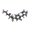  Mass: 322.393 Da / Num. of mol.: 1 / Source method: obtained synthetically / Formula: C18H24F2N2O / Feature type: SUBJECT OF INVESTIGATION Mass: 322.393 Da / Num. of mol.: 1 / Source method: obtained synthetically / Formula: C18H24F2N2O / Feature type: SUBJECT OF INVESTIGATION |
| #4: Chemical | ChemComp-CLR /   Mass: 386.654 Da / Num. of mol.: 1 / Source method: obtained synthetically / Formula: C27H46O Mass: 386.654 Da / Num. of mol.: 1 / Source method: obtained synthetically / Formula: C27H46O |
| #5: Water | ChemComp-HOH /  Mass: 18.015 Da / Num. of mol.: 8 / Source method: isolated from a natural source / Formula: H2O Mass: 18.015 Da / Num. of mol.: 8 / Source method: isolated from a natural source / Formula: H2O |
| Has ligand of interest | Y |
| Has protein modification | Y |
-Experimental details
-Experiment
| Experiment | Method: X-RAY DIFFRACTION / Number of used crystals: 1 |
|---|
- Sample preparation
| Crystal | Density Matthews: 2.21 Å3/Da / Density % sol: 39.44 % |
|---|---|
| Crystal grow | Temperature: 294 K / Method: lipidic cubic phase Details: 100mM sodium cacodylate trihydrate, pH6.4, 90mM sodium citrate, 34% PEG400, 2% Dichloromethane |
-Data collection
| Diffraction | Mean temperature: 100 K / Serial crystal experiment: N |
|---|---|
| Diffraction source | Source: SYNCHROTRON / Site: SSRF / Beamline: BL18U1 / Wavelength: 1.033 Å |
| Detector | Type: DECTRIS PILATUS 6M / Detector: PIXEL / Date: Jun 3, 2021 |
| Radiation | Protocol: SINGLE WAVELENGTH / Monochromatic (M) / Laue (L): M / Scattering type: x-ray |
| Radiation wavelength | Wavelength: 1.033 Å / Relative weight: 1 |
| Reflection | Resolution: 2.6→50 Å / Num. obs: 16071 / % possible obs: 98.5 % / Redundancy: 5.2 % / CC1/2: 0.982 / Rmerge(I) obs: 0.146 / Net I/σ(I): 6.78 |
| Reflection shell | Resolution: 2.6→2.64 Å / Rmerge(I) obs: 0.61 / Num. unique obs: 1448 / CC1/2: 0.522 |
- Processing
| Software |
| |||||||||||||||||||||||||||||||||||||||||||||||||||||||||||||||||||||||||||||||||||||||||||||||||||||||||||||||||||||||||||||||||||||||||||||||||||
|---|---|---|---|---|---|---|---|---|---|---|---|---|---|---|---|---|---|---|---|---|---|---|---|---|---|---|---|---|---|---|---|---|---|---|---|---|---|---|---|---|---|---|---|---|---|---|---|---|---|---|---|---|---|---|---|---|---|---|---|---|---|---|---|---|---|---|---|---|---|---|---|---|---|---|---|---|---|---|---|---|---|---|---|---|---|---|---|---|---|---|---|---|---|---|---|---|---|---|---|---|---|---|---|---|---|---|---|---|---|---|---|---|---|---|---|---|---|---|---|---|---|---|---|---|---|---|---|---|---|---|---|---|---|---|---|---|---|---|---|---|---|---|---|---|---|---|---|---|
| Refinement | Method to determine structure: MOLECULAR REPLACEMENT Starting model: 4u15 Resolution: 2.6→29.896 Å / Cor.coef. Fo:Fc: 0.917 / Cor.coef. Fo:Fc free: 0.864 / SU B: 0.6 / SU ML: 0 / Cross valid method: FREE R-VALUE / ESU R: 0.284 / ESU R Free: 0.353 Details: Hydrogens have been added in their riding positions
| |||||||||||||||||||||||||||||||||||||||||||||||||||||||||||||||||||||||||||||||||||||||||||||||||||||||||||||||||||||||||||||||||||||||||||||||||||
| Solvent computation | Ion probe radii: 0.8 Å / Shrinkage radii: 0.8 Å / VDW probe radii: 1.2 Å / Solvent model: MASK BULK SOLVENT | |||||||||||||||||||||||||||||||||||||||||||||||||||||||||||||||||||||||||||||||||||||||||||||||||||||||||||||||||||||||||||||||||||||||||||||||||||
| Displacement parameters | Biso mean: 44.717 Å2
| |||||||||||||||||||||||||||||||||||||||||||||||||||||||||||||||||||||||||||||||||||||||||||||||||||||||||||||||||||||||||||||||||||||||||||||||||||
| Refinement step | Cycle: LAST / Resolution: 2.6→29.896 Å
| |||||||||||||||||||||||||||||||||||||||||||||||||||||||||||||||||||||||||||||||||||||||||||||||||||||||||||||||||||||||||||||||||||||||||||||||||||
| LS refinement shell |
|
