 Movie
Movie Controller


+ Open data

- Basic information
| Entry | Database: PDB / ID: 7f4r | ||||||
|---|---|---|---|---|---|---|---|
| Title | Crystal structure of MTA1 | ||||||
 Components Components | MT-a70 family protein | ||||||
 Keywords Keywords | DNA BINDING PROTEIN / Protein Complex | ||||||
| Function / homology |  Function and homology information Function and homology informationmRNA m6A methyltransferase / mRNA m(6)A methyltransferase activity / RNA N6-methyladenosine methyltransferase complex / methylation / nucleus Similarity search - Function | ||||||
| Biological species |   Tetrahymena thermophila (eukaryote) Tetrahymena thermophila (eukaryote) | ||||||
| Method |  X-RAY DIFFRACTION / SYNCHROTRON / MOLECULAR REPLACEMENT / Resolution: 1.83 Å X-RAY DIFFRACTION / SYNCHROTRON / MOLECULAR REPLACEMENT / Resolution: 1.83 Å | ||||||
 Authors Authors | Chen, J. / Liu, L. | ||||||
 Citation Citation | Journal: Nat Commun / Year: 2022 Title: Structural basis for MTA1c-mediated DNA N6-adenine methylation Authors: Chen, J. / Hu, R. / Chen, Y. / Lin, X. / Xiang, W. / Chen, H. / Yao, C. / Liu, L. | ||||||
| History |
|
- Structure visualization
| Structure viewer | Molecule: MolmilJmol/JSmol |
|---|
- Downloads & links
-Download
| PDBx/mmCIF format | 7f4r.cif.gz | 179.2 KB | Display | PDBx/mmCIF format |
|---|---|---|---|---|
| PDB format | pdb7f4r.ent.gz | 129.5 KB | Display | PDB format |
| PDBx/mmJSON format | 7f4r.json.gz | Tree view | PDBx/mmJSON format | |
| Others |  Other downloads Other downloads |
-Validation report
| Arichive directory | https://data.pdbj.org/pub/pdb/validation_reports/f4/7f4rftp://data.pdbj.org/pub/pdb/validation_reports/f4/7f4r | HTTPS FTP |
|---|
-Related structure data
| Related structure data | 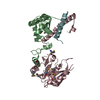 7f4lC  7f4mC 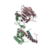 7f4nC 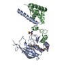 7f4oSC 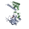 7f4pC 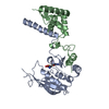 7f4qC 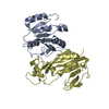 7f4sC 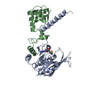 7f4tC S: Starting model for refinement C: citing same article ( |
|---|---|
| Similar structure data |
-Links
 PDBj
PDBj- Assembly
| Deposited unit | 
| ||||||||||||
|---|---|---|---|---|---|---|---|---|---|---|---|---|---|
| 1 | 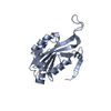
| ||||||||||||
| 2 | 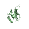
| ||||||||||||
| 3 | 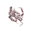
| ||||||||||||
| 4 | 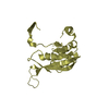
| ||||||||||||
| Unit cell |
|
-Components
| #1: Protein | Mass: 23113.408 Da / Num. of mol.: 4 Source method: isolated from a genetically manipulated source Source: (gene. exp.) Tetrahymena thermophila (strain SB210) (eukaryote)Production host:  #2: Water | ChemComp-HOH / |  Mass: 18.015 Da / Num. of mol.: 541 / Source method: isolated from a natural source / Formula: H2O Mass: 18.015 Da / Num. of mol.: 541 / Source method: isolated from a natural source / Formula: H2O |
|---|
-Experimental details
-Experiment
| Experiment | Method: X-RAY DIFFRACTION / Number of used crystals: 1 |
|---|
- Sample preparation
| Crystal | Density Matthews: 2.22 Å3/Da / Density % sol: 44.66 % |
|---|---|
| Crystal grow | Temperature: 289 K / Method: vapor diffusion, hanging drop / Details: 0.15M potassium sodium tartrate, 18% PEG 3350 |
-Data collection
| Diffraction | Mean temperature: 100 K / Serial crystal experiment: N |
|---|---|
| Diffraction source | Source: SYNCHROTRON / Site: SSRF  / Beamline: BL18U1 / Wavelength: 0.97915 Å / Beamline: BL18U1 / Wavelength: 0.97915 Å |
| Detector | Type: DECTRIS PILATUS3 6M / Detector: PIXEL / Date: May 22, 2021 |
| Radiation | Protocol: SINGLE WAVELENGTH / Monochromatic (M) / Laue (L): M / Scattering type: x-ray |
| Radiation wavelength | Wavelength: 0.97915 Å / Relative weight: 1 |
| Reflection | Resolution: 1.83→50 Å / Num. obs: 54422 / % possible obs: 92.4 % / Redundancy: 5.1 % / Biso Wilson estimate: 18.39 Å2 / Rpim(I) all: 0.049 / Net I/σ(I): 11.79 |
| Reflection shell | Resolution: 1.83→1.86 Å / Mean I/σ(I) obs: 1.43 / Num. unique obs: 3299 / Rpim(I) all: 0.41 |
- Processing
| Software |
| |||||||||||||||||||||||||||||||||||||||||||||||||||||||||||||||||||||||||||||||||||||||||||||||||||||||||
|---|---|---|---|---|---|---|---|---|---|---|---|---|---|---|---|---|---|---|---|---|---|---|---|---|---|---|---|---|---|---|---|---|---|---|---|---|---|---|---|---|---|---|---|---|---|---|---|---|---|---|---|---|---|---|---|---|---|---|---|---|---|---|---|---|---|---|---|---|---|---|---|---|---|---|---|---|---|---|---|---|---|---|---|---|---|---|---|---|---|---|---|---|---|---|---|---|---|---|---|---|---|---|---|---|---|---|
| Refinement | Method to determine structure: MOLECULAR REPLACEMENT Starting model: 7F4O Resolution: 1.83→28.73 Å / SU ML: 0.2199 / Cross valid method: FREE R-VALUE / σ(F): 1.4 / Phase error: 26.4044 Stereochemistry target values: GeoStd + Monomer Library + CDL v1.2
| |||||||||||||||||||||||||||||||||||||||||||||||||||||||||||||||||||||||||||||||||||||||||||||||||||||||||
| Solvent computation | Shrinkage radii: 0.9 Å / VDW probe radii: 1.11 Å / Solvent model: FLAT BULK SOLVENT MODEL | |||||||||||||||||||||||||||||||||||||||||||||||||||||||||||||||||||||||||||||||||||||||||||||||||||||||||
| Displacement parameters | Biso mean: 26.08 Å2 | |||||||||||||||||||||||||||||||||||||||||||||||||||||||||||||||||||||||||||||||||||||||||||||||||||||||||
| Refinement step | Cycle: LAST / Resolution: 1.83→28.73 Å
| |||||||||||||||||||||||||||||||||||||||||||||||||||||||||||||||||||||||||||||||||||||||||||||||||||||||||
| Refine LS restraints |
| |||||||||||||||||||||||||||||||||||||||||||||||||||||||||||||||||||||||||||||||||||||||||||||||||||||||||
| LS refinement shell |
|
