 Movie
Movie Controller

[English] 日本語
 Yorodumi
- PDB-7f1v: Crystal structure of Pseudomonas putida methionine gamma-lyase Q3... -
+ Open data

- Basic information
| Entry | Database: PDB / ID: 7f1v | ||||||
|---|---|---|---|---|---|---|---|
| Title | Crystal structure of Pseudomonas putida methionine gamma-lyase Q349S mutant with L-homocysteine intermediates | ||||||
 Components Components | (L-methionine gamma-lyase) x 2 | ||||||
 Keywords Keywords | LYASE / Pseudomonas putida / methionine gamma-lyase / PLP enzyme | ||||||
| Function / homology |  Function and homology information Function and homology informationhomocysteine desulfhydrase / methionine gamma-lyase / methionine gamma-lyase activity / homocysteine desulfhydrase activity / transsulfuration / : / pyridoxal phosphate binding / cytoplasm Similarity search - Function | ||||||
| Biological species |  Pseudomonas putida (bacteria) Pseudomonas putida (bacteria) | ||||||
| Method |  X-RAY DIFFRACTION / SYNCHROTRON / MOLECULAR REPLACEMENT / Resolution: 2.25 Å X-RAY DIFFRACTION / SYNCHROTRON / MOLECULAR REPLACEMENT / Resolution: 2.25 Å | ||||||
 Authors Authors | Okawa, A. / Handa, H. / Yasuda, E. / Murota, M. / Kudo, D. / Tamura, T. / Shiba, T. / Inagaki, K. | ||||||
 Citation Citation | Journal: J.Biosci.Bioeng. / Year: 2022 Title: Characterization and application of l-methionine gamma-lyase Q349S mutant enzyme with an enhanced activity toward l-homocysteine. Authors: Okawa, A. / Handa, H. / Yasuda, E. / Murota, M. / Kudo, D. / Tamura, T. / Shiba, T. / Inagaki, K. | ||||||
| History |
|
- Structure visualization
| Structure viewer | Molecule: MolmilJmol/JSmol |
|---|
- Downloads & links
-Download
| PDBx/mmCIF format | 7f1v.cif.gz | 303.6 KB | Display | PDBx/mmCIF format |
|---|---|---|---|---|
| PDB format | pdb7f1v.ent.gz | 244.5 KB | Display | PDB format |
| PDBx/mmJSON format | 7f1v.json.gz | Tree view | PDBx/mmJSON format | |
| Others |  Other downloads Other downloads |
-Validation report
| Arichive directory | https://data.pdbj.org/pub/pdb/validation_reports/f1/7f1vftp://data.pdbj.org/pub/pdb/validation_reports/f1/7f1v | HTTPS FTP |
|---|
-Related structure data
| Related structure data |  7f1pC  7f1uC  2o7cS S: Starting model for refinement C: citing same article ( |
|---|---|
| Similar structure data |
-Links
 PDBj
PDBj- Assembly
| Deposited unit | 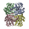
| ||||||||
|---|---|---|---|---|---|---|---|---|---|
| 1 |
| ||||||||
| Unit cell |
|
-Components
| #1: Protein | Mass: 42632.566 Da / Num. of mol.: 2 / Mutation: Q349S Source method: isolated from a genetically manipulated source Source: (gene. exp.) Pseudomonas putida (bacteria) / Gene: mdeA / Production host: References: UniProt: P13254, methionine gamma-lyase, homocysteine desulfhydrase #2: Protein | Mass: 42860.684 Da / Num. of mol.: 2 / Mutation: Q349S Source method: isolated from a genetically manipulated source Source: (gene. exp.) Pseudomonas putida (bacteria) / Gene: mdeA / Production host: References: UniProt: P13254, methionine gamma-lyase, homocysteine desulfhydrase #3: Chemical | 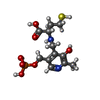  Mass: 366.327 Da / Num. of mol.: 2 / Source method: obtained synthetically / Formula: C12H19N2O7PS / Feature type: SUBJECT OF INVESTIGATION Mass: 366.327 Da / Num. of mol.: 2 / Source method: obtained synthetically / Formula: C12H19N2O7PS / Feature type: SUBJECT OF INVESTIGATION#4: Chemical | ChemComp-HCS / | 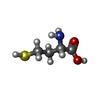  Type: L-peptide linking / Mass: 135.185 Da / Num. of mol.: 1 / Source method: obtained synthetically / Formula: C4H9NO2S / Feature type: SUBJECT OF INVESTIGATION Type: L-peptide linking / Mass: 135.185 Da / Num. of mol.: 1 / Source method: obtained synthetically / Formula: C4H9NO2S / Feature type: SUBJECT OF INVESTIGATION#5: Water | ChemComp-HOH / |  Mass: 18.015 Da / Num. of mol.: 198 / Source method: isolated from a natural source / Formula: H2O Mass: 18.015 Da / Num. of mol.: 198 / Source method: isolated from a natural source / Formula: H2OHas ligand of interest | Y | Has protein modification | Y | |
|---|
-Experimental details
-Experiment
| Experiment | Method: X-RAY DIFFRACTION / Number of used crystals: 1 |
|---|
- Sample preparation
| Crystal | Density Matthews: 2.76 Å3/Da / Density % sol: 55.47 % |
|---|---|
| Crystal grow | Temperature: 293 K / Method: vapor diffusion, hanging drop Details: 0.1 M NA-K PHOSPHATE BUFFER, pH 5.6-5.8, 6-9 % PEG 6000, 0.25 M AMMONIUM SULFATE. 0.5 MM PLP, 0.5%(v/v) 2-mercaptoethanol PH range: 5.6-5.8 |
-Data collection
| Diffraction | Mean temperature: 100 K / Serial crystal experiment: N |
|---|---|
| Diffraction source | Source: SYNCHROTRON / Site: SPring-8  / Beamline: BL44XU / Wavelength: 0.9 Å / Beamline: BL44XU / Wavelength: 0.9 Å |
| Detector | Type: MARMOSAIC 300 mm CCD / Detector: CCD / Date: May 19, 2017 |
| Radiation | Monochromator: Si(111) / Protocol: SINGLE WAVELENGTH / Monochromatic (M) / Laue (L): M / Scattering type: x-ray |
| Radiation wavelength | Wavelength: 0.9 Å / Relative weight: 1 |
| Reflection | Resolution: 2.25→50 Å / Num. obs: 90099 / % possible obs: 97.5 % / Redundancy: 5.2 % / Rmerge(I) obs: 0.045 / Net I/σ(I): 6.5 |
| Reflection shell | Resolution: 2.25→2.29 Å / Redundancy: 4.6 % / Rmerge(I) obs: 0.513 / Mean I/σ(I) obs: 1.3 / Num. unique obs: 4248 / % possible all: 95.4 |
- Processing
| Software |
| ||||||||||||||||||||||||||||||||||||||||||||||||||||||||||||||||||||||||||||||||||||||||||||||||||||||||||||||||||||||||||||||||||||||||||||||||||||||||||||||||||||||||||||||||||||||
|---|---|---|---|---|---|---|---|---|---|---|---|---|---|---|---|---|---|---|---|---|---|---|---|---|---|---|---|---|---|---|---|---|---|---|---|---|---|---|---|---|---|---|---|---|---|---|---|---|---|---|---|---|---|---|---|---|---|---|---|---|---|---|---|---|---|---|---|---|---|---|---|---|---|---|---|---|---|---|---|---|---|---|---|---|---|---|---|---|---|---|---|---|---|---|---|---|---|---|---|---|---|---|---|---|---|---|---|---|---|---|---|---|---|---|---|---|---|---|---|---|---|---|---|---|---|---|---|---|---|---|---|---|---|---|---|---|---|---|---|---|---|---|---|---|---|---|---|---|---|---|---|---|---|---|---|---|---|---|---|---|---|---|---|---|---|---|---|---|---|---|---|---|---|---|---|---|---|---|---|---|---|---|---|
| Refinement | Method to determine structure: MOLECULAR REPLACEMENT Starting model: 2O7C Resolution: 2.25→19.97 Å / Cor.coef. Fo:Fc: 0.947 / Cor.coef. Fo:Fc free: 0.922 / SU B: 6.208 / SU ML: 0.152 / Cross valid method: THROUGHOUT / ESU R: 0.303 / ESU R Free: 0.218 / Stereochemistry target values: MAXIMUM LIKELIHOOD / Details: HYDROGENS HAVE BEEN ADDED IN THE RIDING POSITIONS
| ||||||||||||||||||||||||||||||||||||||||||||||||||||||||||||||||||||||||||||||||||||||||||||||||||||||||||||||||||||||||||||||||||||||||||||||||||||||||||||||||||||||||||||||||||||||
| Solvent computation | Ion probe radii: 0.8 Å / Shrinkage radii: 0.8 Å / VDW probe radii: 1.2 Å / Solvent model: MASK | ||||||||||||||||||||||||||||||||||||||||||||||||||||||||||||||||||||||||||||||||||||||||||||||||||||||||||||||||||||||||||||||||||||||||||||||||||||||||||||||||||||||||||||||||||||||
| Displacement parameters | Biso mean: 38.614 Å2
| ||||||||||||||||||||||||||||||||||||||||||||||||||||||||||||||||||||||||||||||||||||||||||||||||||||||||||||||||||||||||||||||||||||||||||||||||||||||||||||||||||||||||||||||||||||||
| Refinement step | Cycle: 1 / Resolution: 2.25→19.97 Å
| ||||||||||||||||||||||||||||||||||||||||||||||||||||||||||||||||||||||||||||||||||||||||||||||||||||||||||||||||||||||||||||||||||||||||||||||||||||||||||||||||||||||||||||||||||||||
| Refine LS restraints |
|