- PDB-7f1g: BACE2 xaperone complex with N-{3-[(4R,5R,6R)-2-amino-5-fluoro-4,6... -
+
Open data
ID or keywords:
Loading...
-
Basic information
Entry
Database: PDB / ID: 7f1g
Title
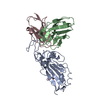
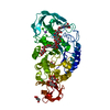
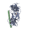
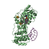
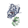
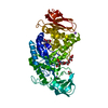
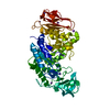
BACE2 xaperone complex with N-{3-[(4R,5R,6R)-2-amino-5-fluoro-4,6-dimethyl-5,6-dihydro-4H-1,3-thiazin-4-yl]-4-fluorophenyl}-2H,3H-[1,4]dioxino[2,3-c]pyridine-7-carboxamide
Components
Beta-secretase 2
XAPERONE
Keywords
HYDROLASE / Beta-secretase 2
Function / homology
Function and homology information
memapsin 1 / negative regulation of amyloid precursor protein biosynthetic process / melanosome organization / melanosome membrane / peptide hormone processing / membrane protein ectodomain proteolysis / amyloid-beta metabolic process / trans-Golgi network / protein processing / glucose homeostasis ...memapsin 1 / negative regulation of amyloid precursor protein biosynthetic process / melanosome organization / melanosome membrane / peptide hormone processing / membrane protein ectodomain proteolysis / amyloid-beta metabolic process / trans-Golgi network / protein processing / glucose homeostasis / aspartic-type endopeptidase activity / endosome / endoplasmic reticulum / Golgi apparatus / proteolysis / membrane / plasma membrane Similarity search - Function
Beta-secretase BACE2 / Beta-secretase BACE / Memapsin-like / Eukaryotic aspartyl protease / Aspartic peptidase A1 family / Peptidase family A1 domain / Peptidase family A1 domain profile. / Aspartic peptidase, active site / Eukaryotic and viral aspartyl proteases active site. / Aspartic peptidase domain superfamily ...Beta-secretase BACE2 / Beta-secretase BACE / Memapsin-like / Eukaryotic aspartyl protease / Aspartic peptidase A1 family / Peptidase family A1 domain / Peptidase family A1 domain profile. / Aspartic peptidase, active site / Eukaryotic and viral aspartyl proteases active site. / Aspartic peptidase domain superfamily / Immunoglobulins / Immunoglobulin-like / Sandwich / Mainly Beta Similarity search - Domain/homology
Beta-secretase2 / Aspartic-like protease 56 kDa / Aspartyl protease 1 / Asp 1 / Beta-site amyloid precursor protein ...Aspartic-like protease 56 kDa / Aspartyl protease 1 / Asp 1 / Beta-site amyloid precursor protein cleaving enzyme 2 / Beta-site APP cleaving enzyme 2 / Down region aspartic protease / DRAP / Memapsin-1 / Membrane-associated aspartic protease 1 / Theta-secretase
Mass: 42105.391 Da / Num. of mol.: 1 Source method: isolated from a genetically manipulated source Source: (gene. exp.) Homo sapiens (human) / Gene: BACE2, AEPLC, ALP56, ASP21, CDA13, UNQ418/PRO852 / Production host: Escherichia coli (E. coli) / References: UniProt: Q9Y5Z0, memapsin 1
#2: Antibody
XAPERONE
Mass: 11968.297 Da / Num. of mol.: 1 Source method: isolated from a genetically manipulated source Source: (gene. exp.) Homo sapiens (human) / Production host: Escherichia coli (E. coli)
In the structure databanks used in Yorodumi, some data are registered as the other names, "COVID-19 virus" and "2019-nCoV". Here are the details of the virus and the list of structure data.
Jan 31, 2019. EMDB accession codes are about to change! (news from PDBe EMDB page)
EMDB accession codes are about to change! (news from PDBe EMDB page)
The allocation of 4 digits for EMDB accession codes will soon come to an end. Whilst these codes will remain in use, new EMDB accession codes will include an additional digit and will expand incrementally as the available range of codes is exhausted. The current 4-digit format prefixed with “EMD-” (i.e. EMD-XXXX) will advance to a 5-digit format (i.e. EMD-XXXXX), and so on. It is currently estimated that the 4-digit codes will be depleted around Spring 2019, at which point the 5-digit format will come into force.
The EM Navigator/Yorodumi systems omit the EMD- prefix.
Related info.:Q: What is EMD? / ID/Accession-code notation in Yorodumi/EM Navigator
Yorodumi is a browser for structure data from EMDB, PDB, SASBDB, etc.
This page is also the successor to EM Navigator detail page, and also detail information page/front-end page for Omokage search.
The word "yorodu" (or yorozu) is an old Japanese word meaning "ten thousand". "mi" (miru) is to see.
Related info.:EMDB / PDB / SASBDB / Comparison of 3 databanks / Yorodumi Search / Aug 31, 2016. New EM Navigator & Yorodumi / Yorodumi Papers / Jmol/JSmol / Function and homology information / Changes in new EM Navigator and Yorodumi
 Movie
Movie Controller

 Yorodumi
 Open data
 Basic information
 Components
 Keywords
 Function and homology information
 Homo sapiens (human)
 X-RAY DIFFRACTION /
  Authors
 Citation
 Structure visualization
 Downloads & links
 Other downloads


 PDBj
PDBj


 Assembly
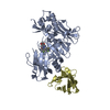

 Mass: 434.460 Da / Num. of mol.: 1 / Source method: obtained synthetically / Formula: C20H20F2N4O3S / Feature type: SUBJECT OF INVESTIGATION
 Mass: 18.015 Da / Num. of mol.: 305 / Source method: isolated from a natural source / Formula: H2O
 Sample preparation
 / Beamline: X06SA / Wavelength: 0.99987 Å
 Processing