 Movie
Movie Controller

[English] 日本語
 Yorodumi
- PDB-7f0c: Crystal structure of capreomycin phosphotransferase in complex wi... -
+ Open data

- Basic information
| Entry | Database: PDB / ID: 7f0c | ||||||
|---|---|---|---|---|---|---|---|
| Title | Crystal structure of capreomycin phosphotransferase in complex with CMN IIA | ||||||
 Components Components |
| ||||||
 Keywords Keywords | TRANSFERASE / Cph / phosphotransferase / capreomycin / resistance | ||||||
| Function / homology |  Function and homology information Function and homology information | ||||||
| Biological species |  Saccharothrix mutabilis subsp. capreolus (bacteria) Saccharothrix mutabilis subsp. capreolus (bacteria) | ||||||
| Method |  X-RAY DIFFRACTION / SYNCHROTRON / MOLECULAR REPLACEMENT / Resolution: 2.07 Å X-RAY DIFFRACTION / SYNCHROTRON / MOLECULAR REPLACEMENT / Resolution: 2.07 Å | ||||||
 Authors Authors | Chang, C.Y. / Pan, Y.C. / Wang, Y.L. / Toh, S.I. | ||||||
| Funding support |  Taiwan, 1items Taiwan, 1items
| ||||||
 Citation Citation | Journal: Acs Chem.Biol. / Year: 2022 Title: Dual-Mechanism Confers Self-Resistance to the Antituberculosis Antibiotic Capreomycin. Authors: Pan, Y.C. / Wang, Y.L. / Toh, S.I. / Hsu, N.S. / Lin, K.H. / Xu, Z. / Huang, S.C. / Wu, T.K. / Li, T.L. / Chang, C.Y. | ||||||
| History |
|
- Structure visualization
| Structure viewer | Molecule: MolmilJmol/JSmol |
|---|
- Downloads & links
-Download
| PDBx/mmCIF format | 7f0c.cif.gz | 72.4 KB | Display | PDBx/mmCIF format |
|---|---|---|---|---|
| PDB format | pdb7f0c.ent.gz | 51.4 KB | Display | PDB format |
| PDBx/mmJSON format | 7f0c.json.gz | Tree view | PDBx/mmJSON format | |
| Others |  Other downloads Other downloads |
-Validation report
| Arichive directory | https://data.pdbj.org/pub/pdb/validation_reports/f0/7f0cftp://data.pdbj.org/pub/pdb/validation_reports/f0/7f0c | HTTPS FTP |
|---|
-Related structure data
| Related structure data |  7f0aSC  7f0bC  7f0fC S: Starting model for refinement C: citing same article ( |
|---|---|
| Similar structure data |
-Links
 PDBj
PDBj

- Assembly
| Deposited unit | 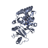
| ||||||||
|---|---|---|---|---|---|---|---|---|---|
| 1 |
| ||||||||
| Unit cell |
|
-Components
| #1: Protein | Mass: 31436.461 Da / Num. of mol.: 1 Source method: isolated from a genetically manipulated source Source: (gene. exp.) Saccharothrix mutabilis subsp. capreolus (bacteria)Gene: cph / Production host: |
|---|---|
| #2: Protein/peptide |   Type: Cyclic peptide / Class: Inhibitor / Mass: 686.723 Da / Num. of mol.: 1 Type: Cyclic peptide / Class: Inhibitor / Mass: 686.723 Da / Num. of mol.: 1Source method: isolated from a genetically manipulated source Source: (gene. exp.) Saccharothrix mutabilis subsp. capreolus (bacteria)Production host: |
| #3: Water | ChemComp-HOH /  Mass: 18.015 Da / Num. of mol.: 131 / Source method: isolated from a natural source / Formula: H2O Mass: 18.015 Da / Num. of mol.: 131 / Source method: isolated from a natural source / Formula: H2O |
| Has ligand of interest | Y |
-Experimental details
-Experiment
| Experiment | Method: X-RAY DIFFRACTION / Number of used crystals: 1 |
|---|
- Sample preparation
| Crystal | Density Matthews: 3.96 Å3/Da / Density % sol: 68.9 % |
|---|---|
| Crystal grow | Temperature: 293 K / Method: vapor diffusion, hanging drop / pH: 7 Details: 1.4 M sodium malonate pH 7.0, 0.1 M Bis-Tris propane pH 7.0, 0.1 M calcium chloride dihydrate |
-Data collection
| Diffraction | Mean temperature: 80 K / Serial crystal experiment: N |
|---|---|
| Diffraction source | Source: SYNCHROTRON / Site: NSRRC / Beamline: BL13B1 / Wavelength: 0.9732 Å |
| Detector | Type: ADSC QUANTUM 210 / Detector: CCD / Date: Dec 17, 2020 |
| Radiation | Protocol: SINGLE WAVELENGTH / Monochromatic (M) / Laue (L): M / Scattering type: x-ray |
| Radiation wavelength | Wavelength: 0.9732 Å / Relative weight: 1 |
| Reflection | Resolution: 2.07→30 Å / Num. obs: 32042 / % possible obs: 99.9 % / Redundancy: 9.6 % / CC1/2: 0.9765 / Net I/σ(I): 58.4 |
| Reflection shell | Resolution: 2.07→2.14 Å / Mean I/σ(I) obs: 3.66 / Num. unique obs: 3156 / CC1/2: 0.888 |
- Processing
| Software |
| ||||||||||||||||||||||||||||||||||||||||||||||||||||||||||||
|---|---|---|---|---|---|---|---|---|---|---|---|---|---|---|---|---|---|---|---|---|---|---|---|---|---|---|---|---|---|---|---|---|---|---|---|---|---|---|---|---|---|---|---|---|---|---|---|---|---|---|---|---|---|---|---|---|---|---|---|---|---|
| Refinement | Method to determine structure: MOLECULAR REPLACEMENT Starting model: 7F0A Resolution: 2.07→25.06 Å / Cor.coef. Fo:Fc: 0.936 / Cor.coef. Fo:Fc free: 0.915 / SU B: 3.978 / SU ML: 0.105 / Cross valid method: THROUGHOUT / σ(F): 0 / ESU R: 0.15 / ESU R Free: 0.147 / Stereochemistry target values: MAXIMUM LIKELIHOOD Details: HYDROGENS HAVE BEEN ADDED IN THE RIDING POSITIONS U VALUES : REFINED INDIVIDUALLY
| ||||||||||||||||||||||||||||||||||||||||||||||||||||||||||||
| Solvent computation | Ion probe radii: 0.8 Å / Shrinkage radii: 0.8 Å / VDW probe radii: 1.2 Å / Solvent model: MASK | ||||||||||||||||||||||||||||||||||||||||||||||||||||||||||||
| Displacement parameters | Biso max: 101.01 Å2 / Biso mean: 32.13 Å2 / Biso min: 13.02 Å2
| ||||||||||||||||||||||||||||||||||||||||||||||||||||||||||||
| Refinement step | Cycle: final / Resolution: 2.07→25.06 Å
| ||||||||||||||||||||||||||||||||||||||||||||||||||||||||||||
| Refine LS restraints |
| ||||||||||||||||||||||||||||||||||||||||||||||||||||||||||||
| LS refinement shell | Resolution: 2.071→2.125 Å / Rfactor Rfree error: 0 / Total num. of bins used: 20
|
