 Movie
Movie Controller


+ Open data

- Basic information
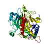
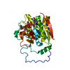
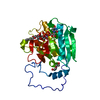
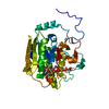
| Entry | Database: PDB / ID: 7ezi | ||||||
|---|---|---|---|---|---|---|---|
| Title | Rice L-galactose dehydrogenase (apo form) | ||||||
 Components Components | L-galactose dehydrogenase | ||||||
 Keywords Keywords | OXIDOREDUCTASE / dehydrogenase | ||||||
| Function / homology |  Function and homology information Function and homology informationL-galactose dehydrogenase activity / L-ascorbic acid biosynthetic process / nucleotide binding / metal ion binding Similarity search - Function | ||||||
| Biological species |  | ||||||
| Method |  X-RAY DIFFRACTION / SYNCHROTRON / MOLECULAR REPLACEMENT / Resolution: 1.2 Å X-RAY DIFFRACTION / SYNCHROTRON / MOLECULAR REPLACEMENT / Resolution: 1.2 Å | ||||||
 Authors Authors | Momma, M. / Fujimoto, Z. | ||||||
 Citation Citation | Journal: To Be Published Title: Crystal structure of rice L-galactose dehydrogenase Authors: Momma, M. / Fujimoto, Z. #1: Journal: Acta Crystallogr Sect F Struct Biol Cryst Commun / Year: 2013 Title: Expression, crystallization and preliminary X-ray analysis of rice L-galactose dehydrogenase. Authors: Momma, M. / Fujimoto, Z. | ||||||
| History |
|
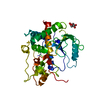
- Structure visualization
| Structure viewer | Molecule: MolmilJmol/JSmol |
|---|
- Downloads & links
-Download
| PDBx/mmCIF format | 7ezi.cif.gz | 140.9 KB | Display | PDBx/mmCIF format |
|---|---|---|---|---|
| PDB format | pdb7ezi.ent.gz | 105.5 KB | Display | PDB format |
| PDBx/mmJSON format | 7ezi.json.gz | Tree view | PDBx/mmJSON format | |
| Others |  Other downloads Other downloads |
-Validation report
| Summary document | 7ezi_validation.pdf.gz | 431.2 KB | Display | wwPDB validaton report |
|---|---|---|---|---|
| Full document | 7ezi_full_validation.pdf.gz | 435 KB | Display | |
| Data in XML | 7ezi_validation.xml.gz | 16.6 KB | Display | |
| Data in CIF | 7ezi_validation.cif.gz | 25.1 KB | Display | |
| Arichive directory | https://data.pdbj.org/pub/pdb/validation_reports/ez/7eziftp://data.pdbj.org/pub/pdb/validation_reports/ez/7ezi | HTTPS FTP |
-Related structure data
| Related structure data |  7ezlC 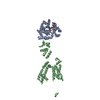 3lutS S: Starting model for refinement C: citing same article ( |
|---|---|
| Similar structure data |


-Links
 PDBj
PDBj
- Assembly
| Deposited unit | 
| ||||||||
|---|---|---|---|---|---|---|---|---|---|
| 1 |
| ||||||||
| Unit cell |
|
-Components
| #1: Protein | Mass: 35299.324 Da / Num. of mol.: 1 Source method: isolated from a genetically manipulated source Source: (gene. exp.) Plasmid: pET-45b / Production host:  |
|---|---|
| #2: Chemical | ChemComp-MG /   Mass: 24.305 Da / Num. of mol.: 1 / Source method: obtained synthetically / Formula: Mg Mass: 24.305 Da / Num. of mol.: 1 / Source method: obtained synthetically / Formula: Mg |
| #3: Water | ChemComp-HOH /  Mass: 18.015 Da / Num. of mol.: 296 / Source method: isolated from a natural source / Formula: H2O Mass: 18.015 Da / Num. of mol.: 296 / Source method: isolated from a natural source / Formula: H2O |
| Has ligand of interest | N |
-Experimental details
-Experiment
| Experiment | Method: X-RAY DIFFRACTION / Number of used crystals: 1 |
|---|
- Sample preparation
| Crystal | Density Matthews: 2.02 Å3/Da / Density % sol: 39 % / Description: rod |
|---|---|
| Crystal grow | Temperature: 293 K / Method: vapor diffusion, sitting drop / pH: 6.5 Details: 0.1M bis-tris pH 6.5, 0.2M MgCl2, 25% poly- ethylene glycol 3350 |
-Data collection
| Diffraction | Mean temperature: 95 K / Serial crystal experiment: N |
|---|---|
| Diffraction source | Source: SYNCHROTRON / Site: Photon Factory  / Beamline: BL-1A / Wavelength: 1 Å / Beamline: BL-1A / Wavelength: 1 Å |
| Detector | Type: ADSC QUANTUM 270 / Detector: CCD / Date: Jan 23, 2012 |
| Radiation | Protocol: SINGLE WAVELENGTH / Monochromatic (M) / Laue (L): M / Scattering type: x-ray |
| Radiation wavelength | Wavelength: 1 Å / Relative weight: 1 |
| Reflection | Resolution: 1.2→100 Å / Num. obs: 87015 / % possible obs: 99.1 % / Redundancy: 7 % / Rmerge(I) obs: 0.077 / Χ2: 1.096 / Net I/σ(I): 17.4 |
| Reflection shell | Resolution: 1.2→1.22 Å / Rmerge(I) obs: 0.611 / Mean I/σ(I) obs: 4.1 / Num. unique obs: 3919 / Χ2: 1.163 |
- Processing
| Software |
| |||||||||||||||||||||||||||||||||||||||||||||||||||||||||||||||||||||||||||||||||||||||||||||||||||||||||||||||||||||||||||||||||||||||||||||||||||
|---|---|---|---|---|---|---|---|---|---|---|---|---|---|---|---|---|---|---|---|---|---|---|---|---|---|---|---|---|---|---|---|---|---|---|---|---|---|---|---|---|---|---|---|---|---|---|---|---|---|---|---|---|---|---|---|---|---|---|---|---|---|---|---|---|---|---|---|---|---|---|---|---|---|---|---|---|---|---|---|---|---|---|---|---|---|---|---|---|---|---|---|---|---|---|---|---|---|---|---|---|---|---|---|---|---|---|---|---|---|---|---|---|---|---|---|---|---|---|---|---|---|---|---|---|---|---|---|---|---|---|---|---|---|---|---|---|---|---|---|---|---|---|---|---|---|---|---|---|
| Refinement | Method to determine structure: MOLECULAR REPLACEMENT Starting model: 3LUT Resolution: 1.2→32.092 Å / Cor.coef. Fo:Fc: 0.968 / Cor.coef. Fo:Fc free: 0.959 / SU B: 1.25 / SU ML: 0.026 / Cross valid method: THROUGHOUT / ESU R: 0.041 / ESU R Free: 0.042 Details: Hydrogens have been used if present in the input file
| |||||||||||||||||||||||||||||||||||||||||||||||||||||||||||||||||||||||||||||||||||||||||||||||||||||||||||||||||||||||||||||||||||||||||||||||||||
| Solvent computation | Ion probe radii: 0.8 Å / Shrinkage radii: 0.8 Å / VDW probe radii: 1.2 Å / Solvent model: MASK BULK SOLVENT | |||||||||||||||||||||||||||||||||||||||||||||||||||||||||||||||||||||||||||||||||||||||||||||||||||||||||||||||||||||||||||||||||||||||||||||||||||
| Displacement parameters | Biso mean: 18.964 Å2
| |||||||||||||||||||||||||||||||||||||||||||||||||||||||||||||||||||||||||||||||||||||||||||||||||||||||||||||||||||||||||||||||||||||||||||||||||||
| Refinement step | Cycle: LAST / Resolution: 1.2→32.092 Å
| |||||||||||||||||||||||||||||||||||||||||||||||||||||||||||||||||||||||||||||||||||||||||||||||||||||||||||||||||||||||||||||||||||||||||||||||||||
| Refine LS restraints |
| |||||||||||||||||||||||||||||||||||||||||||||||||||||||||||||||||||||||||||||||||||||||||||||||||||||||||||||||||||||||||||||||||||||||||||||||||||
| LS refinement shell |
|
