 Movie
Movie Controller


+ Open data

- Basic information
| Entry | Database: PDB / ID: 7eyp | ||||||
|---|---|---|---|---|---|---|---|
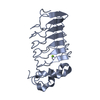
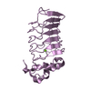
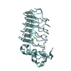
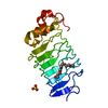
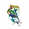
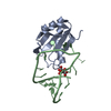
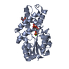
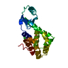
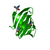
| Title | Crystal structure of Pseudomonas aeruginosa ppnP | ||||||
 Components Components | Pyrimidine/purine nucleoside phosphorylase | ||||||
 Keywords Keywords | HYDROLASE / Pyrimidine / purine / nucleoside phosphorylase | ||||||
| Function / homology |  Function and homology information Function and homology informationpyrimidine-nucleoside phosphorylase / thymidine phosphorylase activity / uridine phosphorylase activity / guanosine phosphorylase activity / purine-nucleoside phosphorylase / purine-nucleoside phosphorylase activity Similarity search - Function | ||||||
| Biological species |   Pseudomonas aeruginosa (bacteria) Pseudomonas aeruginosa (bacteria) | ||||||
| Method |  X-RAY DIFFRACTION / SYNCHROTRON / MOLECULAR REPLACEMENT / Resolution: 1.5 Å X-RAY DIFFRACTION / SYNCHROTRON / MOLECULAR REPLACEMENT / Resolution: 1.5 Å | ||||||
 Authors Authors | Wen, Y. / Wu, B.X. | ||||||
 Citation Citation | Journal: Proteins / Year: 2022 Title: Crystal structures of a new class of pyrimidine/purine nucleoside phosphorylase revealed a Cupin fold. Authors: Wen, Y. / Li, X. / Guo, W. / Wu, B. | ||||||
| History |
|
- Structure visualization
| Structure viewer | Molecule: MolmilJmol/JSmol |
|---|
- Downloads & links
-Download
| PDBx/mmCIF format | 7eyp.cif.gz | 59.3 KB | Display | PDBx/mmCIF format |
|---|---|---|---|---|
| PDB format | pdb7eyp.ent.gz | 41 KB | Display | PDB format |
| PDBx/mmJSON format | 7eyp.json.gz | Tree view | PDBx/mmJSON format | |
| Others |  Other downloads Other downloads |
-Validation report
| Arichive directory | https://data.pdbj.org/pub/pdb/validation_reports/ey/7eypftp://data.pdbj.org/pub/pdb/validation_reports/ey/7eyp | HTTPS FTP |
|---|
-Related structure data
| Related structure data | 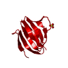 7eyjSC  7eykC  7eylC 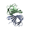 7eymC S: Starting model for refinement C: citing same article ( |
|---|---|
| Similar structure data |
-Links
 PDBj
PDBj- Assembly
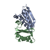
| Deposited unit | 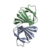
| ||||||||||||
|---|---|---|---|---|---|---|---|---|---|---|---|---|---|
| 1 |
| ||||||||||||
| Unit cell |
|
-Components
| #1: Protein | Mass: 10250.493 Da / Num. of mol.: 2 Source method: isolated from a genetically manipulated source Source: (gene. exp.) Pseudomonas aeruginosa (bacteria) / Gene: ppnP / Production host: #2: Water | ChemComp-HOH / |  Mass: 18.015 Da / Num. of mol.: 307 / Source method: isolated from a natural source / Formula: H2O Mass: 18.015 Da / Num. of mol.: 307 / Source method: isolated from a natural source / Formula: H2O |
|---|
-Experimental details
-Experiment
| Experiment | Method: X-RAY DIFFRACTION / Number of used crystals: 1 |
|---|
- Sample preparation
| Crystal | Density Matthews: 2.58 Å3/Da / Density % sol: 52.28 % |
|---|---|
| Crystal grow | Temperature: 293 K / Method: vapor diffusion, sitting drop / Details: 0.1 M BIS-TRIS pH 5.5, 2.0 M Ammonium sulfate |
-Data collection
| Diffraction | Mean temperature: 100 K / Serial crystal experiment: N |
|---|---|
| Diffraction source | Source: SYNCHROTRON / Site: SSRF  / Beamline: BL18U1 / Wavelength: 0.97915 Å / Beamline: BL18U1 / Wavelength: 0.97915 Å |
| Detector | Type: DECTRIS PILATUS 6M / Detector: PIXEL / Date: May 27, 2021 |
| Radiation | Protocol: SINGLE WAVELENGTH / Monochromatic (M) / Laue (L): M / Scattering type: x-ray |
| Radiation wavelength | Wavelength: 0.97915 Å / Relative weight: 1 |
| Reflection | Resolution: 1.5→30 Å / Num. obs: 33692 / % possible obs: 97.6 % / Redundancy: 19.1 % / CC1/2: 0.989 / CC star: 0.997 / Rmerge(I) obs: 0.103 / Rpim(I) all: 0.024 / Rrim(I) all: 0.105 / Net I/σ(I): 30.875 |
| Reflection shell | Resolution: 1.5→1.55 Å / Redundancy: 19.1 % / Rmerge(I) obs: 0.74 / Mean I/σ(I) obs: 6.75 / Num. unique obs: 3228 / CC1/2: 0.932 / CC star: 0.982 / Rpim(I) all: 0.171 / Rrim(I) all: 0.76 / % possible all: 96.3 |
- Processing
| Software |
| ||||||||||||||||||||||||
|---|---|---|---|---|---|---|---|---|---|---|---|---|---|---|---|---|---|---|---|---|---|---|---|---|---|
| Refinement | Method to determine structure: MOLECULAR REPLACEMENT Starting model: 7EYJ Resolution: 1.5→20 Å / Cross valid method: FREE R-VALUE Stereochemistry target values: GeoStd + Monomer Library + CDL v1.2
| ||||||||||||||||||||||||
| Displacement parameters | Biso mean: 17.32 Å2 | ||||||||||||||||||||||||
| Refinement step | Cycle: LAST / Resolution: 1.5→20 Å
| ||||||||||||||||||||||||
| Refine LS restraints |
|
