 Movie
Movie Controller


+ Open data

- Basic information
| Entry | Database: PDB / ID: 7eva | ||||||
|---|---|---|---|---|---|---|---|
| Title | Structure of molecular chaperone SycE of Yersinia enterocolitica | ||||||
 Components Components | YopE regulator | ||||||
 Keywords Keywords | CHAPERONE / Molecular-chaperone / Yersinia Outer Protein / Type Three secrection chaperone | ||||||
| Function / homology | Type III secretion chaperone SycE / Tir chaperone protein (CesT) family / Tir chaperone protein (CesT) family / protein secretion by the type III secretion system / YopE regulator Function and homology information Function and homology information | ||||||
| Biological species |  Yersinia enterocolitica (bacteria) Yersinia enterocolitica (bacteria) | ||||||
| Method |  X-RAY DIFFRACTION / MOLECULAR REPLACEMENT / Resolution: 2.083 Å X-RAY DIFFRACTION / MOLECULAR REPLACEMENT / Resolution: 2.083 Å | ||||||
 Authors Authors | Kumar, R. / Datta, S. | ||||||
 Citation Citation | Journal: To Be Published Title: Structure of molecular chaperone SycE of Yersinia enterocolitica Authors: Kumar, R. / Datta, S. | ||||||
| History |
|
- Structure visualization
| Structure viewer | Molecule: MolmilJmol/JSmol |
|---|
- Downloads & links
-Download
| PDBx/mmCIF format | 7eva.cif.gz | 40.5 KB | Display | PDBx/mmCIF format |
|---|---|---|---|---|
| PDB format | pdb7eva.ent.gz | 25.4 KB | Display | PDB format |
| PDBx/mmJSON format | 7eva.json.gz | Tree view | PDBx/mmJSON format | |
| Others |  Other downloads Other downloads |
-Validation report
| Arichive directory | https://data.pdbj.org/pub/pdb/validation_reports/ev/7evaftp://data.pdbj.org/pub/pdb/validation_reports/ev/7eva | HTTPS FTP |
|---|
-Related structure data
| Related structure data | 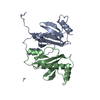 1n5bS S: Starting model for refinement |
|---|---|
| Similar structure data |
-Links
 PDBj
PDBj
- Assembly
| Deposited unit | 
| ||||||||
|---|---|---|---|---|---|---|---|---|---|
| 1 | 
| ||||||||
| Unit cell |
|
-Components
| #1: Protein | Mass: 16914.014 Da / Num. of mol.: 1 Source method: isolated from a genetically manipulated source Source: (gene. exp.) Yersinia enterocolitica (bacteria) / Gene: sycE, yerA, ERS008652_03695, NCTC10938_04290 / Production host: |
|---|---|
| #2: Water | ChemComp-HOH /  Mass: 18.015 Da / Num. of mol.: 75 / Source method: isolated from a natural source / Formula: H2O Mass: 18.015 Da / Num. of mol.: 75 / Source method: isolated from a natural source / Formula: H2O |
-Experimental details
-Experiment
| Experiment | Method: X-RAY DIFFRACTION / Number of used crystals: 1 |
|---|
- Sample preparation
| Crystal | Density Matthews: 1.91 Å3/Da / Density % sol: 35.75 % |
|---|---|
| Crystal grow | Temperature: 298 K / Method: vapor diffusion, sitting drop / pH: 6 Details: 100mM MES pH 6.0, 50-80mM Na-Citrate, 5-10% PEG 3350, 100mM NaCl |
-Data collection
| Diffraction | Mean temperature: 100 K / Serial crystal experiment: N |
|---|---|
| Diffraction source | Source: ROTATING ANODE / Type: Cu FINE FOCUS / Wavelength: 1.5 Å |
| Detector | Type: APEX II CCD / Detector: CCD / Date: Mar 25, 2019 |
| Radiation | Monochromator: M / Protocol: SINGLE WAVELENGTH / Monochromatic (M) / Laue (L): M / Scattering type: x-ray |
| Radiation wavelength | Wavelength: 1.5 Å / Relative weight: 1 |
| Reflection | Resolution: 2.08→25.94 Å / Num. obs: 8342 / % possible obs: 99.7 % / Redundancy: 15.11 % / Rmerge(I) obs: 0.2 / Net I/σ(I): 1.51 |
| Reflection shell | Resolution: 2.08→2.14 Å / Rmerge(I) obs: 0.278 / Num. unique obs: 8342 |
- Processing
| Software |
| ||||||||||||||||||||||||
|---|---|---|---|---|---|---|---|---|---|---|---|---|---|---|---|---|---|---|---|---|---|---|---|---|---|
| Refinement | Method to determine structure: MOLECULAR REPLACEMENT Starting model: 1n5b Resolution: 2.083→25.938 Å / SU ML: 0.13 / Cross valid method: THROUGHOUT / σ(F): 1.34 / Phase error: 22.4 / Stereochemistry target values: ML
| ||||||||||||||||||||||||
| Solvent computation | Shrinkage radii: 0.9 Å / VDW probe radii: 1.11 Å / Solvent model: FLAT BULK SOLVENT MODEL | ||||||||||||||||||||||||
| Displacement parameters | Biso max: 88.81 Å2 / Biso mean: 31.2664 Å2 / Biso min: 12.16 Å2 | ||||||||||||||||||||||||
| Refinement step | Cycle: final / Resolution: 2.083→25.938 Å
| ||||||||||||||||||||||||
| LS refinement shell | Refine-ID: X-RAY DIFFRACTION / Rfactor Rfree error: 0
|
