 Movie
Movie Controller


+ Open data

- Basic information
| Entry | Database: PDB / ID: 7ev5 | ||||||
|---|---|---|---|---|---|---|---|
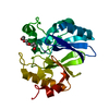
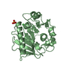
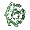
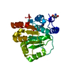
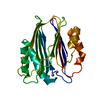
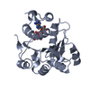
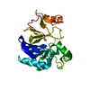
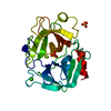
| Title | Crystal structure of BLEG-1 B3 metallo-beta-lactamase | ||||||
 Components Components | Lactamase_B domain-containing protein | ||||||
 Keywords Keywords | HYDROLASE / B3 metallo-beta-lactamase / glyoxalase II / Metallo-hydrolase-like_MBL-fold protein | ||||||
| Function / homology | : / Metallo-beta-lactamase superfamily / Metallo-beta-lactamase superfamily / Metallo-beta-lactamase / Ribonuclease Z/Hydroxyacylglutathione hydrolase-like / metal ion binding / IODIDE ION / Metallo-beta-lactamase domain-containing protein Function and homology information Function and homology information | ||||||
| Biological species |  Bacillus lehensis G1 (bacteria) Bacillus lehensis G1 (bacteria) | ||||||
| Method |  X-RAY DIFFRACTION / MOLECULAR REPLACEMENT / Resolution: 1.44 Å X-RAY DIFFRACTION / MOLECULAR REPLACEMENT / Resolution: 1.44 Å | ||||||
 Authors Authors | Au, S.X. / Muhd Noor, N.D. / Matsumura, H. / Rahman, R.N.Z.R.A. / Normi, Y.M. | ||||||
 Citation Citation | Journal: Int J Mol Sci / Year: 2021 Title: Dual Activity BLEG-1 from Bacillus lehensis G1 Revealed Structural Resemblance to B3 Metallo-beta-Lactamase and Glyoxalase II: An Insight into Its Enzyme Promiscuity and Evolutionary Divergence. Authors: Au, S.X. / Dzulkifly, N.S. / Muhd Noor, N.D. / Matsumura, H. / Raja Abdul Rahman, R.N.Z. / Normi, Y.M. | ||||||
| History |
|
- Structure visualization
| Structure viewer | Molecule: MolmilJmol/JSmol |
|---|
- Downloads & links
-Download
| PDBx/mmCIF format | 7ev5.cif.gz | 64.3 KB | Display | PDBx/mmCIF format |
|---|---|---|---|---|
| PDB format | pdb7ev5.ent.gz | 43.6 KB | Display | PDB format |
| PDBx/mmJSON format | 7ev5.json.gz | Tree view | PDBx/mmJSON format | |
| Others |  Other downloads Other downloads |
-Validation report
| Summary document | 7ev5_validation.pdf.gz | 1.5 MB | Display | wwPDB validaton report |
|---|---|---|---|---|
| Full document | 7ev5_full_validation.pdf.gz | 1.5 MB | Display | |
| Data in XML | 7ev5_validation.xml.gz | 12.4 KB | Display | |
| Data in CIF | 7ev5_validation.cif.gz | 18.2 KB | Display | |
| Arichive directory | https://data.pdbj.org/pub/pdb/validation_reports/ev/7ev5ftp://data.pdbj.org/pub/pdb/validation_reports/ev/7ev5 | HTTPS FTP |
-Related structure data
| Related structure data | 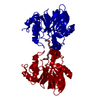 2xf4S S: Starting model for refinement |
|---|---|
| Similar structure data |


-Links
 PDBj
PDBj

- Assembly
| Deposited unit | 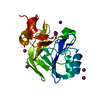
| ||||||||||
|---|---|---|---|---|---|---|---|---|---|---|---|
| 1 |
| ||||||||||
| Unit cell |
|
-Components
| #1: Protein | Mass: 23251.293 Da / Num. of mol.: 1 Source method: isolated from a genetically manipulated source Source: (gene. exp.) Bacillus lehensis G1 (bacteria) / Plasmid: pET-28(b) / Production host: | ||||||
|---|---|---|---|---|---|---|---|
| #2: Chemical |   Mass: 65.409 Da / Num. of mol.: 2 / Source method: obtained synthetically / Formula: Zn / Feature type: SUBJECT OF INVESTIGATION Mass: 65.409 Da / Num. of mol.: 2 / Source method: obtained synthetically / Formula: Zn / Feature type: SUBJECT OF INVESTIGATION#3: Chemical | ChemComp-IOD /   Mass: 126.904 Da / Num. of mol.: 10 / Source method: obtained synthetically / Formula: I Mass: 126.904 Da / Num. of mol.: 10 / Source method: obtained synthetically / Formula: I#4: Water | ChemComp-HOH / |  Mass: 18.015 Da / Num. of mol.: 245 / Source method: isolated from a natural source / Formula: H2O Mass: 18.015 Da / Num. of mol.: 245 / Source method: isolated from a natural source / Formula: H2OHas ligand of interest | Y | |
-Experimental details
-Experiment
| Experiment | Method: X-RAY DIFFRACTION / Number of used crystals: 1 |
|---|
- Sample preparation
| Crystal | Density Matthews: 2.32 Å3/Da / Density % sol: 47 % / Description: Tetragonal |
|---|---|
| Crystal grow | Temperature: 293.15 K / Method: vapor diffusion, sitting drop / Details: NaI, PEG 3350 |
-Data collection
| Diffraction | Mean temperature: 100 K / Serial crystal experiment: N |
|---|---|
| Diffraction source | Source: ROTATING ANODE / Type: RIGAKU MICROMAX-007 / Wavelength: 1.5418 Å |
| Detector | Type: RIGAKU / Detector: CCD / Date: Jul 19, 2019 |
| Radiation | Monochromator: Copper / Protocol: SINGLE WAVELENGTH / Monochromatic (M) / Laue (L): M / Scattering type: x-ray |
| Radiation wavelength | Wavelength: 1.5418 Å / Relative weight: 1 |
| Reflection | Resolution: 1.44→50 Å / Num. obs: 40528 / % possible obs: 98 % / Redundancy: 5.1 % / Rmerge(I) obs: 0.045 / Net I/σ(I): 28.5 |
| Reflection shell | Resolution: 1.44→1.46 Å / Redundancy: 3.1 % / Rmerge(I) obs: 0.254 / Mean I/σ(I) obs: 4 / Num. unique obs: 1991 / CC1/2: 0.899 / % possible all: 97.9 |
- Processing
| Software |
| |||||||||||||||||||||||||||||||||||||||||||||||||||||||||||||||||||||||||||||
|---|---|---|---|---|---|---|---|---|---|---|---|---|---|---|---|---|---|---|---|---|---|---|---|---|---|---|---|---|---|---|---|---|---|---|---|---|---|---|---|---|---|---|---|---|---|---|---|---|---|---|---|---|---|---|---|---|---|---|---|---|---|---|---|---|---|---|---|---|---|---|---|---|---|---|---|---|---|---|
| Refinement | Method to determine structure: MOLECULAR REPLACEMENT Starting model: 2XF4 Resolution: 1.44→30.26 Å / Cross valid method: FREE R-VALUE / Stereochemistry target values: ML
| |||||||||||||||||||||||||||||||||||||||||||||||||||||||||||||||||||||||||||||
| Solvent computation | Solvent model: FLAT BULK SOLVENT MODEL | |||||||||||||||||||||||||||||||||||||||||||||||||||||||||||||||||||||||||||||
| Displacement parameters | Biso mean: 17.3 Å2 | |||||||||||||||||||||||||||||||||||||||||||||||||||||||||||||||||||||||||||||
| Refinement step | Cycle: LAST / Resolution: 1.44→30.26 Å
| |||||||||||||||||||||||||||||||||||||||||||||||||||||||||||||||||||||||||||||
| Refine LS restraints |
| |||||||||||||||||||||||||||||||||||||||||||||||||||||||||||||||||||||||||||||
| LS refinement shell |
|
